Revolutionizing Medical Imaging Technology
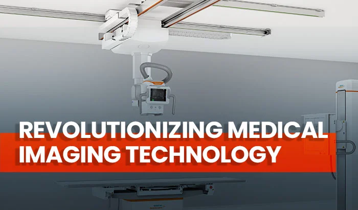
Revolutionizing Medical Imaging Technology: The Impact of Intelligent X-Ray Systems on Workflow and Patient Care
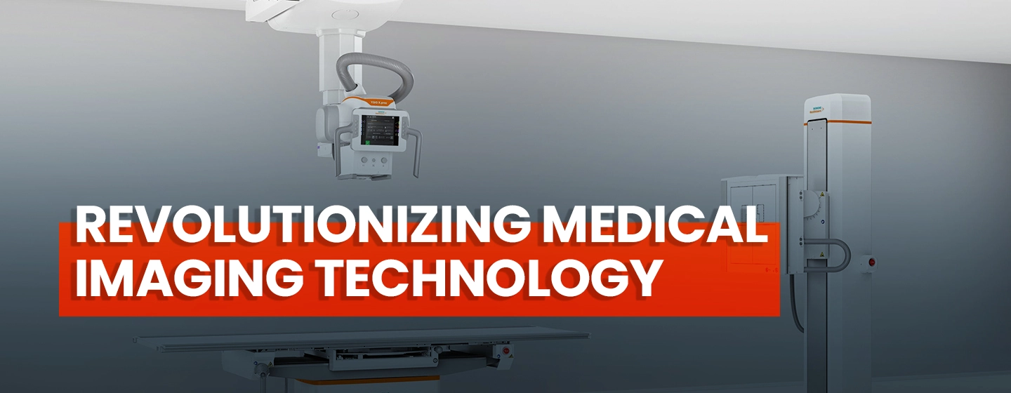

Introduction
Medical imaging technology has evolved impressively over the past decades. While reaching the advanced level of technology depicted in Star Trek—where doctors can instantly diagnose patients by waving a wand—might seem impossible, the rapidly developing field of radiology is making remarkable strides in that direction. This progress is evident with the release of new X-ray intelligence systems. In this article, we will explore the “personal assistant” system that provides automation and support to technologists.

Historical Overview
The history of radiology can be traced back to November 8, 1895, when Wilhelm Roentgen took the first X-ray of a person—specifically, his wife’s hand, now an iconic image.
Roentgen’s X-ray apparatus was widely available, allowing many scientists and physicians to work with and study the medical imaging technology. Many physicians purchased their X-ray machines in the 1910s. Over time, medical imaging technology evolved into a specialized profession distinct from general medicine.

Wilhelm Roentgen (1845-1923), German physicist, received the first Nobel Prize for Physics, in 1901, for his discovery of X-rays in 1895.
Initially, X-ray photographs were created using glass photographic plates, but in 1918, George Eastman introduced film. This innovation resulted in more efficient and possibly less expensive X-ray images. The study of radiology continued into the twentieth century, leading to the founding of the Society of Radiographers in 1920.
Medical imaging technology advancements improved the speed and usefulness of X-rays, and their applications expanded. More safety laws and measures were realized and implemented throughout the 1940s and 1950s. X-rays became more valuable in treatment and diagnosis, helping address more serious medical issues.
The computer was a significant technological advancement in the 1970s and 1980s. These breakthroughs were game-changers, as computerized X-rays provided dramatically improved imaging and information.
The 1980s and 1990s also saw the birth of teleradiology as a new medical imaging technology asset, which enabled radiologists, physicians, and healthcare facilities to easily and quickly communicate and share patient images via computers, scanners, and other equipment. This innovation led to incredible collaboration between radiologists and physicians, resulting in overall better patient care.
Need for Development
As X-ray technologists, we walk hundreds of miles at work each year. On average, a technologist works 40 hours a week, 52 weeks a year, and takes approximately 9.5 minutes and 52 steps per exam, with each step averaging 2.4 feet. This amounts to walking 311 miles a year—quite a workout!
But it’s not just the steps that contribute to fatigue and burnout. Click, push, pull, and walk! On a typical day, an X-ray technologist must perform all these actions to complete a single workflow. Whether it’s dragging the machine from one location to another or walking back and forth to position the patient, the Bucky, or the tube, these tasks add up over time. This can lead to negative outcomes, such as increased exam time, physical exertion for the technologist, errors, and stress.
X-ray imaging often serves as the entry point to diagnostic imaging, accounting for 60% of all imaging studies conducted. As a result, X-ray technologists, radiology administrators, and radiologists face an ever-increasing caseload. The daily challenges of heavy lifting, repetitive motions, uneasy patients, and long hours result in more than 70% of technologists suffering work-related injuries. Variability in patient positioning and exam setup can lead to additional radiation dose and “repeat and reject” rates of up to 25%, necessitating retakes of images.
Given the stress and pressure of keeping the radiology department running smoothly, a collaborative effort between technologists, radiologists, and administrators is essential to increase efficiency and enhance outcomes. However, traditional X-ray systems often fail to meet today’s expectations and frequently obstruct these efforts due to extensive workflow processes, imprecise operation, complicated patient setup, poor image quality, physically demanding movements, and limited clinical applications.
To address these challenges, imaging device manufacturers such as Siemens, Agfa, and GE have developed the “personal assistant” system as a new medical imaging technology. This system reduces the number of miles a technologist walks over a year, ensuring that every component of care uplifts each individual who interacts with the system to provide the best patient care possible.
Description of the Advances
In October 2020, Siemens Healthineers announced FDA clearance of the YSIO X.pree, a ceiling-mounted radiography system with the MyExam Companion intelligent user interface. Agfa HealthCare (Mortsel, Belgium) launched its SmartXR digital radiography (DR) portfolio at RSNA 2020, and GE Healthcare followed in September 2021 with the introduction of the Definium Tempo, a new fixed, overhead tube suspension digital X-ray system.
These new machines use a unique combination of hardware and AI-powered software to lighten radiographers’ workloads and provide image acquisition support. They are designed to be a “personal assistant” to radiologists and technologists, leveraging automation to reduce workflow burdens and help radiology departments deliver the best patient care possible.
Let’s explore the characteristics of these machines and how they will make our work easier.
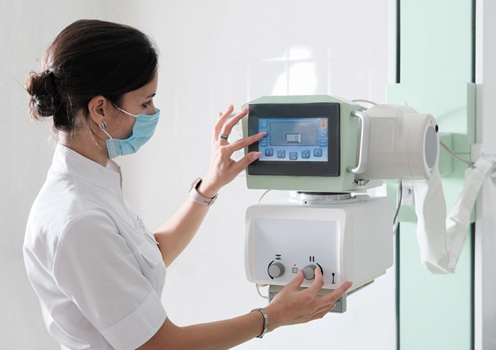
The tube-mounted console, which functions similarly to an in-room command center, provides all the functionality required for patient selection, protocol selection, technique modification, and positioning setup. This allows the technologist to complete all exam setup and positioning work without having to walk back and forth to the acquisition workstation outside the room.
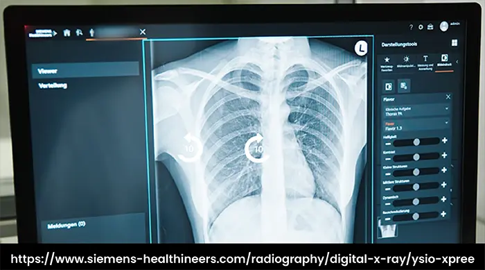
The technologist is guided through the exam procedure by the intelligent user interface. It offers proactive information to assist technologists of all skill levels in navigating a radiography procedure. The interface combines available patient data, such as gender and age, with other user or machine-observable, patient-specific information to determine the best acquisition and reconstruction parameters for each patient and radiography procedure. These innovations improve the efficiency of the imaging process.
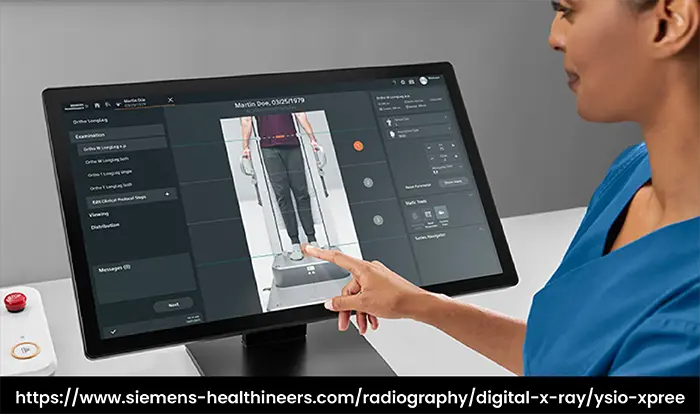
The 3-D camera in the system helps to speed up the clinical workflow. With its Virtual Collimation feature, technologists can accurately adjust the size and shape of the X-ray beam via the touchscreen monitor, reducing the volume of irradiated patient tissue.
The system automatically moves the wall stand, table, and OTS to custom predefined positions based on the exam protocol, and the preset auto-positioning is optimized for speed. These movements can be initiated by the OTS console, a remote control, or at the acquisition workstation.
To address “repeat and reject” rates, which can negatively impact day-to-day clinical operations, the system includes features that deliver consistent images and help reduce variability in patient positioning and image quality. Enabled by live streaming video and a 3D depth camera, the Intelligent Workflow Suite combines computer vision, video analytics, and precision engineering to provide a collection of workflow enhancement tools to address positioning errors, poor image quality due to incorrect patient habitus selection, and ambiguities during image reading due to unique conditions.
Position Assist, Technique Assist, and Patient Snapshot are included in the Intelligent Workflow Suite to boost patient satisfaction by eliminating image retakes and providing more consistent images to increase technologists’ productivity.
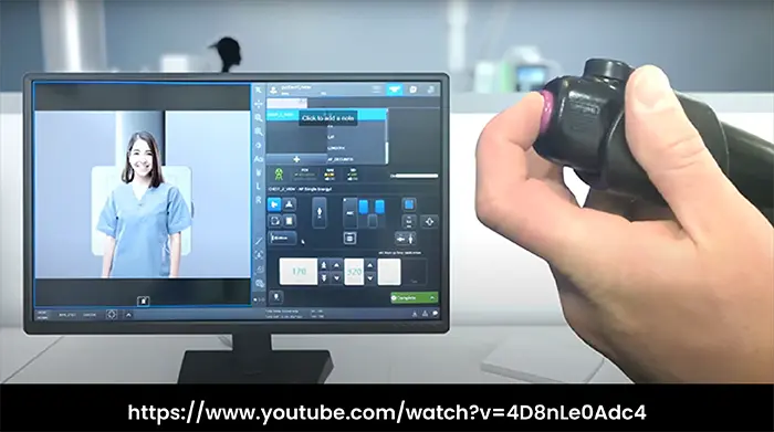
Shorter preparation periods for orthopedic multi-image acquisitions are possible thanks to the camera-based Smart Virtual Ortho functionality. With Smart Virtual Ortho, the technologist can adjust exposure parameters and select the field of view on the touchscreen while viewing a live camera image of the patient, making long-leg and full-spine examinations faster and easier to complete. The camera’s Auto Thorax Collimation function accelerates thorax examination workflows by assessing the patient’s body contour and modifying the collimator blades in less than half a second.
The system’s image processing database is designed to provide image consistency. Intelligent dynamic range optimization can be used by technologists to set optimal visual contrasts. Intelligent noise removal eliminates image noise and allows for low-dose settings for all clinical applications, while grid-less acquisition for free exposures reduces the requirement for additional patient radiation dose. AI-based cropping is intended to produce consistently well-cropped images.
To assist technologists, manufacturers used automation to reduce patient positioning time, physical workload, errors, and image retakes through a user-centered design with simple features that lower the number of workflow steps and actions necessary. Auto Positioning, Auto Centering, Auto and Reverse Tracking, and Auto Angulation automate the positioning of system components to maximize overall ergonomic operation for the technologist while also contributing to an improved patient experience.
“I’ve been a technologist for over 17 years, and using this system has been unlike anything I’ve experienced before—it’s like having a person inside the room with you at all times, like you’re never working alone. It’s like a second technologist,” said Nadia Dorson, X-ray Technologist at North Central Bronx Hospital. “When you’re in front of your patient, you’re able to double-check the patient’s information, which is really important for setting up the exam properly. The camera provides real-time information on the patient, so you can make necessary adjustments. And everything is as light as a feather, even down to the grid, so I’m able to function better and avoid some of the day-to-day wear and tear I’ve experienced in the past.”
Indeed, the “personal assistant” systems are intended to collaborate with the technologist to operate quickly and provide consistent, high-quality images while allowing for more quality time with the patient.
To boost your knowledge, we recommend you to enjoy the interactive experience proposed by Agfa on this link:
https://medimg.agfa.com/
Advantages
The X-Ray intelligence systems offer numerous advantages:
- Ease the physical burden: The ergonomic design of GE and Siemens systems prioritizes the well-being of the team. Features like the tiltable multitouch monitor, remote control, and wireless barcode reader reduce walking distances. The flat table design and low table height enable comfortable patient positioning, while the fully automated system swiftly moves into position at the touch of a button, minimizing physical strain on technologists
- Simplify workflow: By minimizing workflow steps, automatically moving system components, and intelligently confirming information, these systems speed up exams, eliminate retakes, and reduce pressure on technologists
- Improve diagnostic confidence: These systems assist radiologists by delivering consistently high-quality images through industry-leading detectors, advanced image processing, and innovative applications like image pasting and dual-energy subtraction
- Easy and undisrupted operation: The “personal assistant” feature makes it easier for technologists to interact with the system and their patients. The system includes a camera that allows technologists to keep the patient in focus at all times, providing confidence using the 3D Camera. It also assists in identifying movements and promotes high-quality outcomes. The 3D Camera delivers a live patient image to the workstation, enabling virtual collimation, auto thorax collimation, and smart virtual ortho
- Provide optimal support during examinations: The system improves consistency across different users with the Positioning Guide, which can be customized to meet the team’s needs and increase standardization. Favorite Tools provide immediate access to the most relevant tools for the team, enabling smoother, faster, more standardized post-processing in all clinical exams
- Reduce user-dependent variations: Sound diagnoses require consistent images while keeping patient dose to a minimum. Dose Adaptions allow the team to modify dose settings to its standards, using patient-size brackets established in the Patient Size Adapter (S, M, L, XL). Smart Virtual Ortho allows for accurate collimation, helping users quickly determine if they can perform an exam with fewer images. Dose adjustments per clinical images can further lower dose
All these benefits contribute to the main goal of medical staff: better patient care.
“The system has helped strengthen the role of the technologist as a member of the larger health care team,” shares Dr. Orlando Ortiz, Radiologist at North Central Bronx Hospital. “All of a sudden, the team becomes that much more effective, because as you strengthen each link, you get a better chain overall. This has a direct impact on the patient, because when technologists spend less time looking at screens and more time being with the patient, patients feel more reassured and less anxious. The medical imaging technology, in this case, has gotten it right; we’re actually doing the right thing for the patient because it enables the technologist to focus on one of the reasons they wanted to become a technologist in the first place: to take care of people.” (2)
Conclusion
The YSIO X.pree, SmartXR DR, and Definium Tempo systems represent a significant advancement in intelligent X-ray imaging, offering radiology departments unprecedented levels of intuitive operation for consistent X-ray images and potentially faster workflows. The innovative medical imaging technology in these systems has shown a direct positive impact on patients and staff members.
As the field of medical imaging technologies continues to advance, this path to intelligent imaging extends further with the introduction of the Revolution Ascend by GE Healthcare on September 21, 2021. This new system promises to bring the same level of innovation and improvement to CT scanning, further enhancing the capabilities of radiology departments and their ability to provide high-quality patient care.
References
- https://www.xray.com.au/
- https://www.gehealthcare.com/
- https://www.itnonline.com/
- https://www.gehealthcare.com/
- https://www.siemens-healthineers.com/
- https://www.itnonline.com/
- https://www.ge.com/
- https://www.medimaging.net/
Disclaimer: The information provided on this website is intended to provide useful information to radiologic technologists. This information should not replace information provided by state, federal, or professional regulatory and authoritative bodies in the radiological technology industry. While Medical Professionals strives to always provide up-to-date and accurate information, laws, regulations, statutes, rules, and requirements may vary from one state to another and may change. Use of this information is entirely voluntary, and users should always refer to official regulatory bodies before acting on information. Users assume the entire risk as to the results of using the information provided, and in no event shall Medical Professionals be held liable for any direct, consequential, incidental or indirect damages suffered in the course of using the information provided. Medical Professionals hereby disclaims any responsibility for the consequences of any action(s) taken by any user as a result of using the information provided. Users hereby agree not to take action against, or seek to hold, or hold liable, Medical Professionals for the user’s use of the information provided.
