Radiation Protection in PET/CT

Radiation Protection in PET/CT


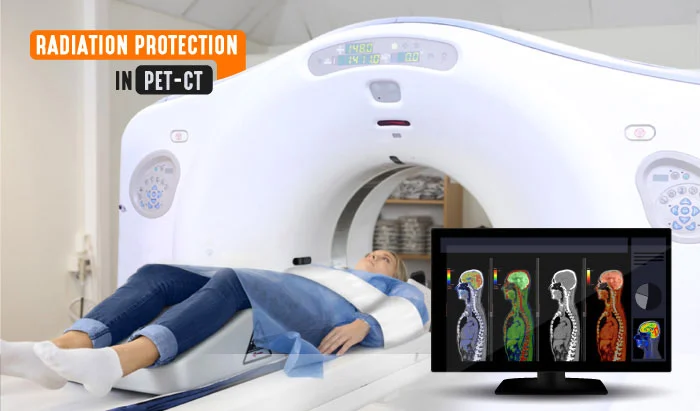
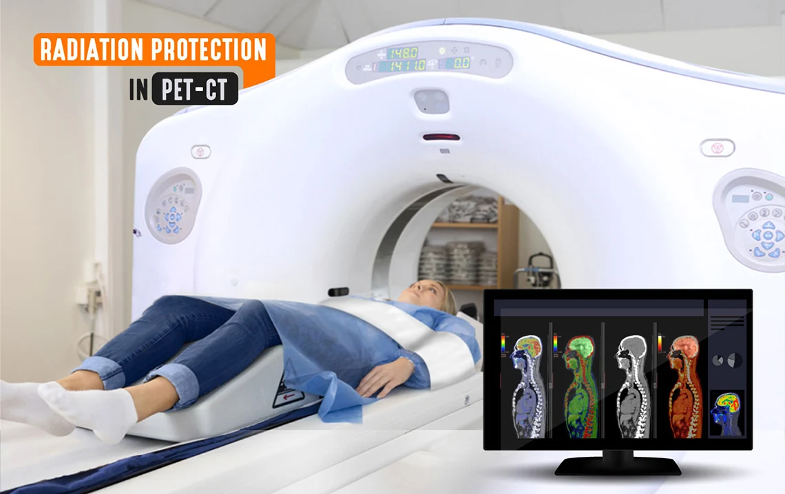
Did you know that a CT scan of the abdomen and pelvis exposes an individual to about 10 mSv, while a PET/CT exposes an individual to about 25 mSv of radiation? This is equal to about 8 years of average background radiation exposure. PET is used to examine metabolic processes within the body and is very important in the detection of CA and CA metastasis. The single joint imaging device of PET/CT affords medical professionals with the ability to detect the presence of abnormally high regions of glucose metabolism, which may indicate cancer spread or metastasis, and obtain detailed information in reference to the location and extent of these lesions. In this article, we will discuss radiation doses in computerized tomography (CT) and positron emission tomography (PET), as well as the PET/CT radiation protection measures necessary to keep both the patient dose and the occupational dose to a minimum.
Sources of Radiation in PET/CT
There are two sources of radiation contributing the radiation dose for patients having a PET/CT. The first source is internal. For a PET scan, patients are injected with 18F-FDG, which is a positron emitter and results in the production of high energy radiation. The second source is external, when the patient then has the CT scan.

There are also two possible sources of radiation contributing to the occupational radiation dose in PET/CT. The first possible source of radiation exposure is when the healthcare professional is handling radiopharmaceuticals such as 18F-FDG. Exposure can occur throughout the handling process, which encompasses dose preparation, transport of the syringe into the injection room, the injection of the patient, measuring the residual activity, and handling the radioactive waste post-injection. The second source of radiation is the medical professional’s close contact with the patient after the administration of 18F-FDG, which includes contact occurring during the uptake stage, while escorting the patient to the scanning room, during the scanning stage, and also during the release stage.

PET/CT Radiation Protection Principles and Procedures
Radiation Protection Principles
- Justification refers to doing more good than harm when deciding if radiation use is necessary
- Optimization refers to using the minimal amount of time during radiation exposure, the largest amount of distance from the radiation source, and shielding whenever possible
- Dose limitations are for occupational radiation in order to manage workers’ exposure. They include, but are not limited to, a maximum radiation effective dose of 20 mSv per year, 500 mSv per year to the skin, hands, and feet, and 20 mSv per year to the lens of the eye
Procedures to Protect Medical Personnel from PET/CT Radiation
In diagnostic imaging, the only concern is the radiation emanating from the X-ray machine. But in PET/CT, there are multiple radiation concerns: the scatter radiation coming from the CT scanner and the high energy annihilation photons emanating in all directions from both the patient having the PET/CT and the patient in the nearby injection or preparation room waiting to be scanned. Each 18F nuclear transformation by positron decay yields two highly penetrating 511 kEv photons that are unable to be shielded by an ordinary lead apron. The thickness necessary to attenuate this type of high energy radiation by 50% is about .5 cm. (.2 inches), and adequate shielding at close distances could require up to 2.5 cm or an inch of lead.

- Use of the inverse square law to maximize distance between the patient and personnel, as the absorbed dose decreases substantially with increased distance between the radiation source, in this case the patient, and personnel
- The appropriate use at all times of PET/CT radiation protection, including lead shielding, walls, and the gantry
- Use of electronic personal dosimeters to serve as a constant reminder of exposure
- The importance of using shielding dispensing units, syringe shields, and carriers
- Reducing occupational radiation dose to technologists and staff by standing at either the end of the scanning bed or to the side of the gantry in order to reduce radiation dose

Procedures to Reduce Patient PET/CT Radiation Dose
Radiation exposure to the PET/CT patient can be greatly reduced by modifying the acquisition parameters for CT. Because radiation effects are cumulative in nature, repeat procedures are to be avoided as much as possible. This requires knowledgeable CT technologists to perform the procedures. Because children are more sensitive to ionizing radiation, it is important to justify all pediatric examinations, and those examinations should be performed with due optimization. A patient must also carefully be interviewed to assess the likelihood of pregnancy in order to minimize any unintentional radiation exposure to an embryo or fetus. Clearly visible notices should be placed throughout the area informing patients of the need and importance of disclosing any possibility of pregnancy before receiving any radioactive material by the physician or technologist.

In order to keep both patient dose and the occupational radiation doses to an absolute minimum or ALARA, “as low as reasonably achievable,” all of the PET/CT radiation protection techniques mentioned within this article are essential and should be practiced diligently by the PET/CT technologist at all times. If you want to learn more about radiation protection in different modalities, check out our Radiation Protection in Radiology CE course (8 CE credits).
References
Peet, D., Morton, R., Hussein, M., Alsafi, K., & Spyrou, N. (2012, May). Radiation protection in fixed PET/CT facilities–design and operation. Retrieved January 13, 2021, from https://www.ncbi.nlm.nih.gov/
Hosono, M., Komeya, Y., Im, S., Hamahata, K., Okada, S., Yamamoto, K., Tsuichiya, N. (2007, May 01). Radiation safety in PET/CT practices: Justification and optimization for patients and personnel. Retrieved January 13, 2021, from https://jnm.snmjournals.org/
Alice, S. S. (2018). Radiation protection: In medical radiography (8th ed.). St Louis, Missouri: Elsevier.


