X-Ray Positioning Guide: Toes
X-Ray Radiographic Positioning Guide: Toes


This little piggy went to market. This little piggy went home. This little piggy got smashed into a filing cabinet and went weeeee… all the way to the radiologic technologist for imaging. This article is intended as a quick X-ray positioning guide for radiologic technologists and addresses radiographic positioning to obtain high-quality X-rays of patients’ toes.
It may be that it’s been awhile since you’ve needed to X-ray of all of a patient’s toes, or it may be that you are looking for guidance on the technical parameters to improve the quality of your X-rays. Whatever your toe X-ray needs, this guide should help.
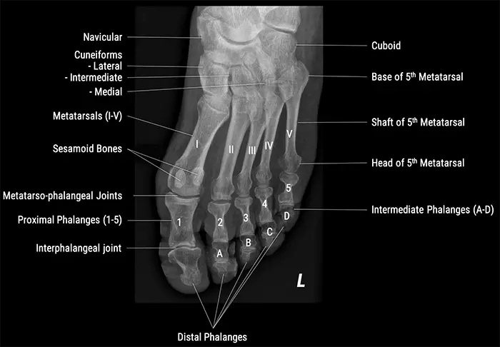
X-Ray Positioning Guide: Anatomy of the Toes
Before we move into the X-ray positioning guide proper, we’ll start with the basics: the anatomical names of the toes (which are, we know, much less adorable than just pointing and saying “this little piggy”).
X-Ray Positioning Guide: Radiographic Positioning
The guide below includes the technical parameters, patient positioning, tube and centering point, criteria for success, and indications for each view of the toes. Click on the collapsible menu to see detailed information for each of the radiographic positioning views below.
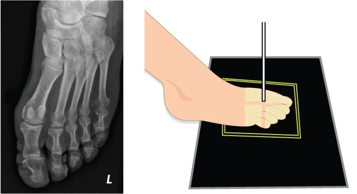
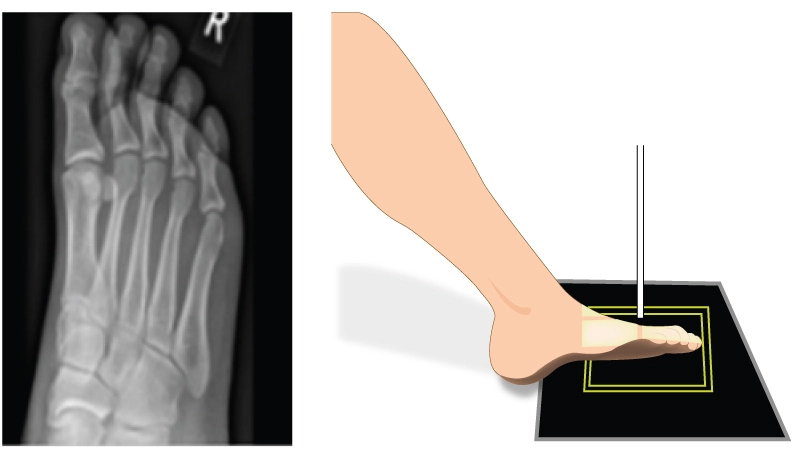
AP Dorsoplantar View
- Orientation: Portrait
- Detector:
- Cassette size: 18 cm x 24 cm: 45-47 kV – 6-8 mAs
- Or Digital Panel Detector: 49-52 kV – 1.6-2 mAs
- SID = 100 cm
- The patient can be either supine or sitting upright
- The knee should be flexed so the plantar surface of the foot makes contact with the detector
- AP projection
- Centering point: x-ray beam centered to the metatarsophalangeal joint in question; the beam is perpendicular to the detector
- On X-ray, toe(s) of interest should be seen independently with no overlapping of soft tissues
- No rotation should be present; the shafts of the phalanges and metatarsals appear equal on either side
- Fractures, dislocation, foreign body, some pathologies such as osteoarthritis and gout
Lateral View
- Orientation: Portrait
- Detector:
- Cassette size: 18 cm x 24 cm: 45-47 kV – 6-8 mAs
- Or Digital Panel Detector: 49-52 kV – 1.6-2 mAs
- SID = 100 cm
- The patient may be supine or upright depending on comfort
- Depending on the toe of interest and to reduce magnification, the affected leg may be medially (1st to 3rd toes) or laterally (4th and 5th toes) rotated
- Mediolateral/Lateromedial projection
- Centering point: interphalangeal joint of 1st digit and proximal interphalangeal joint of 2nd to 5th digit
- The condyles of the proximal phalanx should be superimposed
- Increased concavity on the plantar surface of the proximal phalanx is demonstrated
- Fractures, dislocation, foreign body, some pathologies such as osteoarthritis and gout
Oblique View
- Orientation: Portrait
- Detector:
- Cassette size: 18 cm x 24 cm: 45-47 kV – 6-8 mAs
- Or Digital Panel Detector: 49-52 kV – 1.6-2 mAs
- SID = 100 cm
- The patient may be supine or upright
- The affected leg must be flexed enough that the plantar aspect of the foot is resting on the image receptor
- The foot is medially rotated until the plantar surface sits at a 45° angle to the image receptor
- AP oblique projection
- Centering point: x-ray beam centered to the metatarsophalangeal joint in question; the beam will be perpendicular to the image receptor
- Superimposition is evident at the bases of the of 1st and 2nd metatarsals
- There is no superimposition of the 3rd to 5th metatarsal
- Fractures, dislocation, foreign body, some pathologies such as osteoarthritis and gout
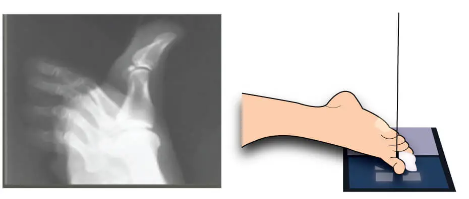
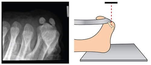
Sesamoid View: Holly Method
- Orientation: Portrait
- Detector:
- Cassette size: 18 cm x 24 cm: 45-47 kV – 6-8 mAs
- Or Digital Panel Detector: 49-52 kV – 1.6-2 mAs
- SID = 100 cm
- The patient may be supine or sitting upright with their leg straightened on the table
- The foot is in dorsiflexion
- The toes are pulled back toward the patient
- Axial projection
- Centering point: base of the first metatarsal
- Both of the sesamoid bones are free from the superimposition
- The extent of injury to sesamoid bones
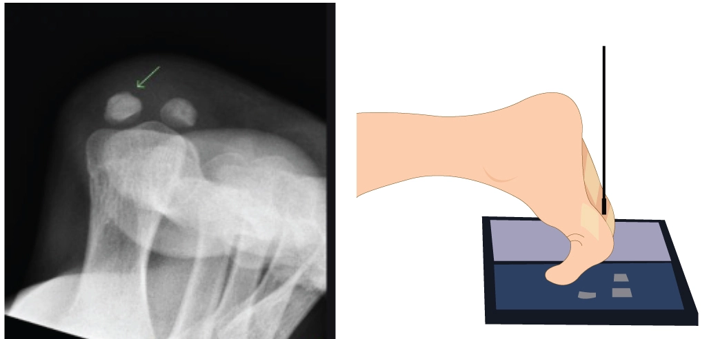
Sesamoid View: Lewis
- Orientation: Portrait
- Detector:
- Cassette size: 18 cm x 24 cm: 45-47 kV – 6-8 mAs
- Or Digital Panel Detector: 49-52 kV – 1.6-2 mAs
- SID = 100 cm
- The patient in prone position
- Pillow are used for patient comfort
- The foot is in dorsiflexion
- Toe on the table
- Axial projection
- Centering point: base of the first metatarsal
- Both of the sesamoid bones are free from the superimposition
- The extent of injury to sesamoid bones
If you found this guide to x-ray positioning for toes useful and would like to suggest other guides to radiographic positioning that you’d like to see added to our Reference Guides, feel free to contact us with your suggestions.

