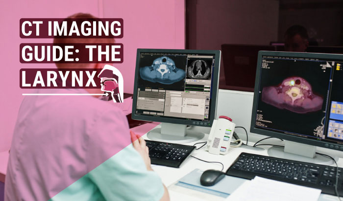
CT Imaging Guide: The Larynx

CT Imaging Guide: The Larynx


Computed Tomography (CT) scanning can be a valuable tool in assessing trauma to and the many different pathologies of the laryngeal space. This article is part of our series on Computed Tomography (CT), and addresses the larynx and laryngeal space, including anatomy, pathological findings, and CT scanning protocols specific to the larynx.
Laryngeal Anatomy – Larynx CT
The larynx is located between the third and sixth cervical vertebra in the anterior compartment of the neck. It is suspended from the hyoid bone and is continuous inferiorly with the trachea. It opens superiorly at the laryngeal segment of the pharynx, situated between the pharynx and the first tracheal ring. The larynx is a cartilaginous skeleton with ligaments and muscles that move and stabilize it along with a mucous membrane.

Source: https://www.health.harvard.edu/
Serving as a conduit for the passage of air between the pharynx and the trachea, the larynx regulates airflow through its lumen and protects the lower airway during swallowing. It contains the vocal cords for producing sounds—known as phonation—and functions as a voice box.

Source: https://www.criticalcarepractitioner.co.uk/
- Thyroid cartilage
- Cricoid cartilage
- Epiglottis
- Arytenoid cartilages
- Corniculate cartilages
- Cuneiform cartilages
The thyroid cartilage, cricoid cartilage, and epiglottis are unpaired, while the arytenoid, corniculate, and cuneiform cartilages are paired. The thyroid, the largest segment, is a protective shield which surrounds the anterior section of the larynx and meets in the middle at the protrusion known as the laryngeal prominence, or, as it is more commonly known, the “Adam’s apple.”
The internal space of the larynx is wide superiorly and inferiorly but narrows in the middle, forming a section named glottis. The glottis is the portion of the laryngeal cavity formed by the four vocal folds and the opening between the folds. The other two sections are the supraglottic, and infraglottic.

Source: https://www.semanticscholar.org/
CT Imaging of the Larynx
Multidetector Computed Tomography (MDCT) is usually the first choice for cross-sectional imaging in the diagnostic workup of laryngeal lesions. The advantages of MDCT is its ability for thin slices which provide high spatial resolution. This, in turn, allows for higher quality multiplanar reformation.
When imaging cancer of the larynx with CT, technologists should perform contrast-enhanced helical CT scanning from the C1 vertebral body to the thoracic inlet, with the section plane parallel to the true vocal cords or the hyoid bone. The section thickness should not be greater than 3 mm, with patients instructed to breathe quietly. With current multidetector-row CT scanners, collimation can be 1 mm, with the larynx imaged in just a few seconds, providing near-isometric Z-axis resolution with minimal motion artifacts.
Using soft tissue or bone window settings, the exact location, vascularization, extension of lesions, and their involvement of the laryngeal skeleton can be evaluated easily. When determining an appropriate biopsy site, especially in submucosal lesions that have overlying normal mucosa, MDCT can play a vital role. Both MDCT and magnetic resonance imaging (MRI) can also provide the assessment of cervical lymph node involvement.
MRI provides more accurate and detailed information in evaluating submucosal spaces, subglottic, and tongue base infiltration. Because of its high soft tissue contrast resolution, it has the potential for better tissue characterization.
Laryngeal Pathology – Larynx CT
Squamous cell carcinoma, the most common laryngeal neoplasm which accounts for approximately 95% of all malignant neoplasm of the larynx, is closely correlated with smoking. Head and neck tumors occur 6 times more often in cigarette smokers than in nonsmokers. However, there are many other benign and malignant tumors and inflammatory diseases that can affect the larynx. Imaging studies may offer clues in the diagnosis of non-squamous cell laryngeal lesions. Previous clinical history along with current clinical and imaging findings should be evaluated together in the management of laryngeal lesions.

(A) 34 year-old woman with laryngeal paraganglioma. Right pre-epiglottic well defined enhanced mass (arrows) is seen in contrast-enhanced CT
(B) Contrast-enhanced T1 weighted turbo spin echo spectral fat saturation inversion recovery (T1 TSE SPIR).
(C) Axial 68Gallium-DOTA-peptide PET/CT fusion image.
(D) Intense uptake by the right laryngeal paraganglioma similar with synchronous left carotid body paraganglioma.

(A) 70 year-old man with laryngeal chondroma. Contrast-enhanced CT image shows expansile mass arising from cricoid cartilage. Chondroid calcifications are seen within the mass.
(B) Coronal T2 weighted TSE image shows high signal intensity transglottic mass.
(C) Endoscopy reveals bilateral edema of the mucosa of the arytenoid cartilages and a large submucosal mass.
External blunt trauma to the neck can be caused by motor vehicle accidents, assaults, sports-related trauma, and strangulation. These are the most common causes of laryngeal fractures. Penetrating trauma due to stab wounds or gunshots is the second leading cause of these types of fractures. The images below show an isolated longitudinal fracture of the thyroid cartilage due to a motor vehicle accident.

(Creative Commons Attribution -ShareAlike 4.0 International License)
Axial (A) and coronal (B) 2D with bone windows shows a paramedian, longitudinal fracture of the thyroid cartilage as noted by the arrows with major soft tissue emphysema. (C) 3D VR (anterior view) of the thyroid cartilage (yellow) and airways (blue) depicts the cranio-caudal etent of the fracture line (arrow) more pricesly.
Laryngeal Trauma
Performed under general anesthesia, and usually as an ambulatory procedure with the patient returning home the same day, micro-laryngoscopy takes about one hour. The surgeon works in collaboration with the anesthesiologist during this procedure. The pain following this surgery is minor, rarely requiring prescription pain medications.

- Vocal cord stripping to remove the superficial layers of the vocal cords
- Laser surgery to remove the entire vocal cords
- Partial laryngectomy removing only the diseased portion of the larynx and sparing as much of the vocal cords as possible
Recap of CT Protocols for the Larynx
- Contrast-enhanced helical CT from the C1 vertebral body to the thoracic inlet
- Section plane parallel to the true vocal cords or the hyoid bone
- Section thickness should not be greater than 3 mm
- Patients should be instructed to breathe quietly
- With current multidetector-row CT scanners, collimation can be 1 mm
References
Allen E, Minutello K, Murcek BW. Anatomy, Head and Neck, Larynx Recurrent Laryngeal Nerve. [Updated 2020 Jul 27]. In: StatPearls [Internet]. Treasure Island (FL): StatPearls Publishing; 2020 Jan-. [Figure, Recurrent laryngeal nerves. Image courtesy S Bhimji MD] Available from: https://www.ncbi.nlm.nih.gov/
Becker M, Burkhardt K, Dulguerov P, Allal A. Imaging of the larynx and hypopharynx. Eur J Radiol. 2008 Jun. 66(3):460-79. [Medline]
Doğan S, Vural A, Kahriman G, İmamoğlu H, Abdülrezzak Ü, Öztürk M. Non-squamous cell carcinoma diseases of the larynx: clinical and imaging findings. Braz J Otorhinolaryngol. 2020;86:468–82.


