Exposure Index, Deviation Index, and Optimal Radiographic Imaging

Exposure Index and indicator, Deviation Index, and Optimal Radiographic Imaging


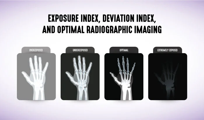
What is the difference between exposure index and deviation index in digital imaging, and how can you know if you have an optimal radiographic image? This article aims to answer both questions for rad techs to support you in acquiring optimal radiographic images. We will cover the definitions and concepts of exposure index, exposure indicator and deviation index and how both relate to producing optimal images that are neither underexposed, nor overexposed.
The Challenge of Optimal Image Acquisition
Various manufacturers utilize different image receptor exposure indices or exposure indicators in order for a radiologic technologist to be able to discern between a radiographic image that is optimal and radiographic images that may be overexposed or underexposed. With the wide dynamic range that digital imaging allows, it is extremely difficult for radiologic technologists to determine if the resultant image is optimal due to the look-up table re-scaling the image to match the stored histogram in the computer. Image processing will adjust the resultant radiographic image and still produce the ultimate gray scale, but the trade-off is a lack of accurate visual feedback regarding exposure to the image receptor. With this in mind, rad techs face significant challenges in producing an optimal radiographic image at a reasonably low patient dose.
Exposure Index and Deviation Index
Exposure index (EI) measures the amount of x-ray photons that actually reach the image receptor in the relative image region. The deviation index (DI) measures the difference between how many photons should be reaching the image receptor in the relative image region for that particular study and the amount of x-ray photons that actually do reach the image receptor in the relative image region. The EI depends on the examination type, image processing, and exposure. At the same kVp setting, doubling the mAs settings doubles the EI value in direct exposure systems, and halving the mAs settings will half the EI value in a direct imaging radiographic system.
Equipment manufacturers are all heading in the direction of utilizing deviation indices rather than exposure indicators, and the American College of Radiology (ACR) is in the process of developing a method to track and record all radiography studies as they do currently with computed tomography.
Exposure Index Values and Image Quality
With direct digital exposure systems, if the EI value is below the manufacturer’s normal range, the radiograph is underexposed, causing a decrease in radiographic quality. These types of images are said to have “quantum mottle” or “noise” and may appear grainy. If the EI value is above the manufacturer’s normal range, then the radiograph is overexposed. In this case, dose creep, or the administration of excessive radiation to the patient, is an issue. There are still some indirect radiographic systems in use with EI values indicating the opposite indications. With these systems, if the EI is lower than the normal range, then the radiograph is overexposed, and if the value is higher than the normal range, then the radiograph is underexposed.
Deviation Index Values and Image Quality
The DI is an indication as to whether or not the radiographic technique used is appropriate for the specific body part and for an optimal presentation of the anatomy of interest, proper image brightness and contrast, with an acceptable signal-to-noise ratio throughout the resultant radiographic image. While each manufacturer of radiographic equipment has varied EI ranges, DI values consistently range on a continuum of -3 to +3, with a certain range in between being acceptable according to the equipment’s calibration. A DI of +1 indicates an overexposure of 25% more than the desired exposure to the image receptor, while a DI of -1 indicates an underexposure of 20% less than desired exposure to the IR. DI values of +3 and -3 indicate two times more or less than the desired exposure to the image receptor, respectively.
Target Exposure Index
EIT or the target exposure index, is the reference exposure obtained when an image is properly exposed. This exposure indicator is set by the equipment manufacturer or by the imaging center and is dependent on the body part, view, procedure, and imaging receptor. DI quantifies how much the actual EI varies from the EIT as stated above. In an ideal scenario, when the EI is the same as the EIT , the DI will be 0.
An objective EIT for common examinations needs to be established on the basis of image quality. Quality assurance programs, which utilize exported measures that are recorded in DICOM structured reports, are also needed. These reports can then be monitored and systemic trends can also be identified and corrected. The ACR has already established this monitoring system for CT and is investigating implementing a Dose Index Registry for digital radiography that can also be used to establish national benchmarks for image receptor exposure.
Final Thoughts and Further Learning
Digital radiographic systems produce images with consistent density and contrast regardless of the exposure factors selected by the radiologic technologist. For this reason, exposure indicators were developed in order to guide the technologist and assist them in selecting the proper exposure factor that will produce an optimal radiographic image that is neither overexposed, nor underexposed.
We hope this article has helped you gain a better understanding of the exposure index and deviation index and how they relate to the acquisition of high quality radiographs. If you want to learn more about how to improve the quality of the images you acquire, check out our Data Acquisition and Processing course (1.5 CE Credits for ARRT® certification).


