Digital Subtraction Angiography and CT Angiography

Objectives:
After reading this article you should be able to:
- Review the anatomy and physiology of the Brain
- Explain the process of subtraction in CT angiography
- Compare digital subtraction angiography vs CT angiography
Introduction
Subtraction is a post-processing technique that is used to eliminate high-density structures, such as bone, from CT images. A subtraction dataset is obtained by subtracting a non-contrast acquisition known as a mask image from a contrast-enhanced image. To accomplish this, accurate registration of the two datasets is essential. In contrast to digital subtraction angiography, CT angiography is 3D-based and therefore more challenging. In addition, the minimal time delay between the non-contrast and contrast acquisitions can introduce differences between scans due to 3D-based patient motion or vascular pulsation. To be able to overcome these challenges, a dedicated registration algorithm is applied, and this registration algorithm matches the position of bony structures and calcifications in the body on the mask image to the contrast scan before subtraction. As a result, subtracted image data are obtained from which high-density structures have been removed and which can be used for evaluation in conjunction with the conventional contrast-enhanced images. This article will review the anatomy and physiology of the brain, explain the process of subtraction in CT angiography, and compare digital subtraction angiography vs CT angiography.
Cerebral anatomy
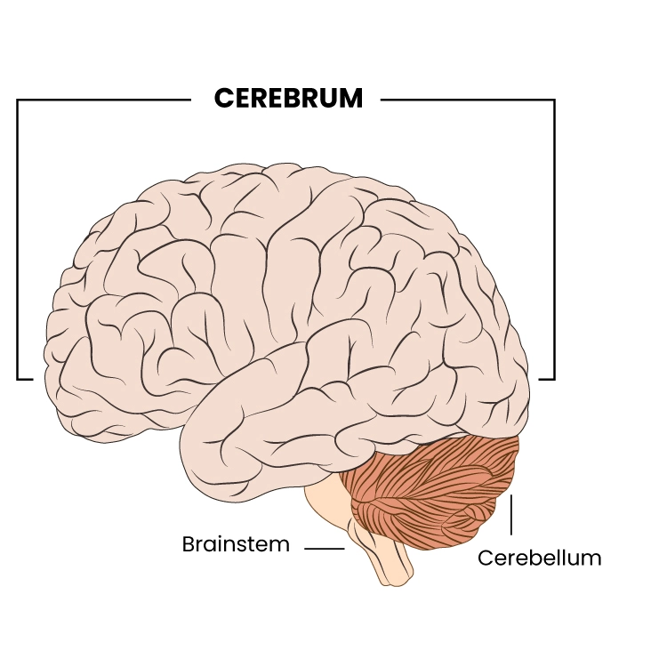
The brain is a complex organ that controls thought, memory, emotion, touch, motor skills, vision, breathing, temperature, hunger, and every process that regulates our body. Together, the brain and spinal cord that extend from it make up the central nervous system or CNS.
Weighing about 3 pounds in the average adult, the brain is about 60% fat. The remaining 40% is a combination of water, protein, carbohydrates, and salts. The brain itself is not a muscle but has blood vessels and nerves, including neurons and glial cells.
Gray and white matter are two different regions of the central nervous system. In the brain, gray matter refers to the darker, outer portion, while white matter describes the lighter, inner section underneath. Each region serves a different role with gray matter primarily responsible for processing and interpreting information, while white matter transmits that information to other parts of the nervous system.
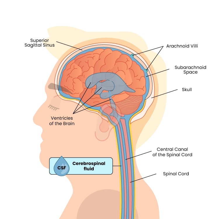
Deep in the brain are four open areas with passageways between them. They also open into the central spinal canal and the area beneath the arachnoid layer of the meninges. The ventricles manufacture cerebrospinal fluid, or CSF, a watery fluid that circulates in and around the ventricles and the spinal cord, and between the meninges. CSF surrounds and cushions the spinal cord and brain washes out waste and impurities, and delivers nutrients.
Blood Supply
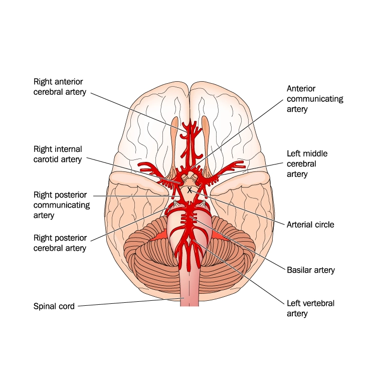
Two sets of blood vessels supply blood and oxygen to the brain: the vertebral arteries and the carotid arteries. The external carotid arteries extend up the sides of your neck and are where you can feel your pulse when you touch the area with your fingertips. The internal carotid arteries branch into the skull and circulate blood to the front part of the brain. (“Brain Anatomy and How the Brain Works | Johns Hopkins Medicine”)
The vertebral arteries follow the spinal column into the skull, where they join at the brainstem and form the basilar artery, which supplies blood to the rear portions of the brain.
“The Circle of Willis, a loop of blood vessels near the bottom of the brain that connects major arteries, circulates blood from the front of the brain to the back and helps the arterial systems communicate with one another.” (“Brain Anatomy and How the Brain Works | Johns Hopkins Medicine”)
Substraction CT Angiography
In subtraction CT angiography (sCTA), a non-contrast CT acquisition is subtracted from a contrast-enhanced CTA acquisition. sCTA has been shown to produce comparable results to digital subtraction angiography (DSA) in the detection and classification of intracranial aneurysms. sCTA is advantageous over DSA because of its non-invasive nature, shorter examination time, and lower costs. Small aneurysms and aneurysms next to bony structures are more accurately detected in sCTA as compared to conventional CTA. (“Ultra-high-resolution subtraction CT angiography in the follow-up of …”)
Ultra High Resolution (UHR) Cerebral Subtraction CTA
Image acquisition with Ultra High-Resolution CT (UHRCT) is comparable to the standard 80-row CT scanning. A typical protocol for cerebral sCTA with UHRCT consists of two consecutive high-resolution scans, one pre-contrast and one contrast-enhanced, using orbital synchronization. Scanning should be performed in super high-resolution mode. Typically, a bolus of 50-ml contrast medium is injected with an injection rate of 5 ml/s which is followed by a saline flush. Following acquisition, images are automatically reconstructed with a 0.25-mm thickness, a 0.25-mm interval, and a 240-mm FOV.
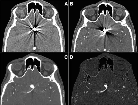
The pre-and post-contrast scans are automatically registered and subtracted, supplying a subtraction CT dataset in addition to the standard reconstructions. An example illustrating the impact of the available post-processing techniques (joint MAR and MBIR followed by subtraction) on metal artifact reduction and image quality in a patient with aneurysmal coil embolization is provided in Fig. below.
https://media.springernature.com
Impact of post-processing on image quality and metal artifact reduction. a Non-enhanced CT and b CT angiography with pronounced scattering artifacts of a coil-treated anterior communicating artery aneurysm. c CT angiography with metal artifact reduction (MAR) and model-based iterative reconstruction (MBIR) provide substantial artifact reduction. d Subtraction CT angiography with MAR and MBIR filtering provides further artifact reduction and results in adequate image quality for diagnostic evaluation
Digital Subtraction Angiography vs CT Angiography – A SUMMARY
Digital Subtraction Angiography and CT Angiography are both imaging techniques used to visualize blood vessels and diagnose vascular conditions. However, they differ in terms of the technology used and the information they provide. Here is a comparison between digital subtraction angiography and CT angiography:
Technology- How are digital subtraction angiography and CT angiography different?
- DSA: DSA is an X-ray-based technique. It involves the injection of a contrast agent and the acquisition of X-ray images before and after the injection. Subtraction imaging is then performed to visualize the blood vessels.
- CTA: CTA is a form of computed tomography (CT) scanning. It uses X-rays and a contrast agent to generate cross-sectional images of the blood vessels. A CT scanner rotates around the body, capturing multiple X-ray images from different angles. These images are reconstructed to create detailed 3D images.
Image Quality- Is One Better Than the Other?
- DSA: DSA provides excellent image quality specifically for visualizing blood vessels. Using subtraction imaging enhances the visibility of blood vessels while minimizing the interference of other structures.
- CTA: CTA supplies high-resolution 3D images that offer detailed information about the blood vessels and surrounding anatomy. It can also visualize soft tissues and organs in addition to the blood vessels.
Radiation Exposure-Do digital subtraction angiography and CT angiography differ in doses to the patient?
- DSA: DSA involves the use of X-rays, which expose the patient to a certain amount of radiation. However, advancements in technology have reduced the radiation dose associated with DSA.
- CTA: CTA also uses X-rays, and the radiation exposure can vary depending on the scanning protocol and the specific equipment used. Newer CT scanners employ dose-reduction techniques to minimize radiation exposure.
Procedure and Workflow- Invasive vs Non-Invasive digital subtraction angiography and CT angiography
- DSA: DSA is an invasive procedure that requires the insertion of a catheter into a blood vessel, usually through the groin or arm. It is performed in an interventional radiology suite or angiography suite. The contrast agent is directly injected into the blood vessels.
- CTA: CTA is a non-invasive procedure that involves the injection of a contrast agent intravenously. The patient lies on a CT scanner table, which moves through the scanner to acquire images. It is typically performed in a radiology department.
Applications
- DSA: DSA is commonly used to assess the patency and integrity of blood vessels, detect blockages, diagnose aneurysms, and guide interventional procedures such as angioplasty or embolization.
- CTA: CTA is used for various vascular evaluations, including assessing the coronary arteries for coronary artery disease, evaluating the pulmonary arteries for pulmonary embolism, examining the aorta and peripheral arteries for stenosis or aneurysms, and evaluating the cerebral arteries for stroke or vascular malformations.
The choice between digital subtraction angiography and CT angiography depends on the specific clinical situation, the anatomical region of interest, and the preference of the healthcare provider. Each technique has its advantages and limitations, and the selection is made based on the individual patient’s needs and the desired diagnostic information.
