CT Contrast Agents, Extravasation, and Treatment: Rad Tech Fundamentals


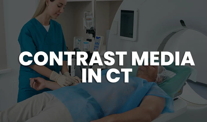
Patient care is the most important component of any CT examination. This includes communication with the patient, the procedures that are completed prior to starting an exam, during the exam and finally following the CT examination. Contrast agents are used in CT to help highlight blood vessels and enhance tissue structures. The purpose of a contrast-enhanced CT (CECT) scan is to find pathologies by enhancing the contrast between a lesion and the normal surrounding structures. Follow this article to learn about CT contrast extravasation treatment and injection and agents.
Radiologic technologists’ role in a CECT scan procedure is integral to providing optimal patient care and obtaining the right diagnosis. The purpose of this article (which is part of our CT basics course) is to provide you with a comprehensive guide to the possible complications you might encounter while using contrast agents in CT.

Contrast Agent Injection
There are important considerations you must take when administering IV contrast. Foremost, It is imperative to draw the contrast agent using a sterile syringe to prevent infection. Also, use a power injector in order to administer the contrast under slow perfusion. If the patient already has an IV in place, use a different site to inject the contrast media to avoid any local complications at the injection site.
A good glomerular filtration rate is important for contrast toxicity prevention. When radiographic contrast media are injected intravenously or intra-arterially, they pass from the vascular compartment into the extracellular space through capillaries. They are then eliminated almost entirely by glomerular filtration. Check the patient’s estimated glomerular filtration rate (eGFR) before administering any contrast agent to prevent any possible toxicity.
Phases of a CECT scan
A non-enhanced CT helps in detecting calcifications, fat in tumors, and fat-stranding as seen in inflammations like appendicitis, diverticulitis, omental infarction, etc. On the other hand, contrast-enhanced CT has three phases, and each one helps in detecting specific structures and pathologies. CECT phases are based on where the contrast media is located during imaging.
Arterial Phase
The first phase is the arterial phase and it occurs 15-20 sec after bolus tracking. Structures that get their blood supply from the arteries will show optimal enhancement.
Hepatic Phase
Later comes the Hepatic or late portal phase at 50-60 sec after bolus. Although the hepatic phase is the most accurate term, most people use the term “late portal phase”. In this phase, the liver parenchyma enhances due to the blood supply from the portal vein. At that point, some enhancement of the hepatic veins should be noticed.
Delayed Phase
Lastly, the delayed phase is usually 6-10 minutes after bolus. Sometimes called the “wash-out phase” or “equilibrium phase”. There, contrast media diffuse to all abdominal structures except for fibrotic tissue which will become relatively dense compared to normal tissue. This makes the delayed phase optimal for visualizing abdominal structures and hidden abdominal ailments.
Contrast Extravasation
Various side effects might occur during a CECT procedure. One of the most common side effects is extravasation. IV infiltration occurs when fluid infuses into the tissues surrounding the venipuncture site. This sometimes happens when the tip of the catheter slips out of the vein. Either the catheter passes through the wall of the vein, or the blood vessel wall allows part of the fluid to infuse into the surrounding tissue.
Extravasation occurs when there is accidental infiltration of a vesicant that can cause blistering or seepage of contrast into the surrounding IV site. Vesicants can cause tissue destruction and/or blistering. Pain can result at the IV site and along the vein and may or may not cause inflammation. Extravasation is an unwanted occurrence that can cause irreversible local injuries. It results in tissue sloughing, pain, loss of mobility in the extremity, and infection.
Extravasation of CT contrast agent into upper extremity subcutaneous tissue is a relatively frequent complication of injection. Treat upper extremity extravasation by elevating and massaging the affected limb and use warm compresses to dull the pain.

Contrast Agent Concerns
Non-renal acute adverse reactions to iodinated contrast agents for CT include flushing, nausea, arm pain, pruritus, vomiting, headache, and urticaria. These symptoms may be felt approximately 30 seconds after the beginning of the injection. The peak sensations are usually felt after 50 seconds (in the complete portal phase), then slow down after 1 minute.
While such discomforts are not serious, they still constitute legitimate patient concerns. Inform the patient of the likelihood to encounter such discomforts and assure them that these feelings are perfectly normal and will pass soon. Below are three common patient concerns that rad techs encounter during a CECT scan:
- Flushing: CT contrast agents cause vasodilation, which will be expressed as a flushed feeling by the patient. Most patients will notice a very warm feeling that spreads throughout their body for about 20 seconds during and after the injection. This can startle patients who are poorly informed, making it difficult for them to cooperate with breath-hold commands, and thus decrease the quality of the examination.
- Urination: Additionally, flushing is often concentrated around the groin area because of its large blood supply. This can cause patients to feel like they are passing urine. Such sensations are very common and usually go away quickly (10–20 seconds).
- Metallic Taste: When the contrast diffuses into the salivary glands, the patient might be startled by its metallic taste. Reassure the patient beforehand that this is a normal sensation.
Allergies
Whereas the above symptoms are mild and command little medical concern, the allergic symptoms we will discuss hereafter require immediate medical attention. Allergy symptoms may appear within minutes of contrast injection and progress to more serious symptoms, including itching of the eyes or face. Varying degrees of mouth, throat, and tongue swelling can make breathing and swallowing difficult. Other allergy symptoms include hives (urticaria), abdominal pain and cramps, vomiting, uncontrolled diarrhea, and mental confusion or dizziness.

- Urticaria: Urticaria, also known as hives, is an outbreak of swollen, pale red bumps or plaques on the skin that appear suddenly. It is a result of the body’s reaction to the contrast agent used in certain CT examinations.
- Pseudo-allergy: Reactions to contrast media are not considered a true allergy – no allergic antibody exists that causes the reaction. Rather, contrast media act directly to release histamine and other chemicals from mast cells. The severity of the reaction depends on the iodine concentration: the higher the iodine concentration, the greater the risk of an adverse reaction. Rad Techs should keep an eye out for a rare but dangerous pseudo-allergy that can prevent a patient from breathing – Quincke’s angioedema. Signs and symptoms in patients suffering from Quincke’s angioedema may include the following: swelling of the face (eyelids, lips), tongue, hands, and feet. More severe signs and symptoms include throat tightness, voice changes, breathing trouble, and most concerning a swollen uvula.

- Anaphylaxis: The terms “anaphylaxis” and “anaphylactic shock” are often used to mean the same thing. Both refer to a severe allergic reaction. Shock occurs when blood pressure drops so low that the cells and organs seize to receive enough oxygen. Anaphylactic shock is a type of shock that’s caused by a severe allergic reaction.
CECT scan procedure risks
A CECT scan isn’t considered a risky procedure. However, there are certain patients at increased risk from CECT scan complications. Apply great care when injecting iodinated contrast into these high-risk patients.
Contrast-Induced Nephropathy
Contrast-induced nephropathy (CIN), which is the deterioration of kidney function as a result of iodine-based contrast, can have serious effects. Increased morbidity, as well as mortality rates, have been demonstrated in patients at risk. Those patients include heart patients, diabetics, the elderly, and those with known poor renal function and abnormal lab values.

The most important prophylactic measure for CIN is hydration. Prior to the day of the exam, encourage all patients to have oral hydration that includes water, coffee, and tea. After the examination, instruct the patients to drink at least 5 (8-ounce) glasses of water or until their urine becomes clear. This will help flush the contrast medicine from their body. Certain precautions should be taken with pregnant and breastfeeding women as well:
- Pregnancy: CT examinations may be contraindicated in patients who are pregnant or suspected of being pregnant unless the procedure benefits out way the risks to the mother and the fetus. When possible an ultrasound should be used. If not possible, or in the case of trauma, a CT scan can be performed with the fewest slices and lowest doses possible to reduce the fetal dose. Always consult a radiologist before the such an examination.

- Breastfeeding: Generally, iodine from iodinated contrast media (either oral or injectable types) is distributed in very small quantities into breast milk. Based on kinetic studies, it is unlikely that these agents will reach therapeutic levels in breast milk, and no adverse effects in infants have been observed following maternal use of iodinated contrast agents in CT. Both the American Academy of Pediatrics (AAP) and the American College of Radiology (ACR) consider that the use of iodinated contrast agent is compatible with breastfeeding.
- Pacemakers and Defibrillators: Unlike magnetic resonance imaging, a CT scan can be done even if a patient has a pacemaker or cardioverter defibrillator. Although it is possible that CT x-rays directly irradiating the electronics of implantable pacemakers or ICDs can cause electronic interference, the probability that this interference can cause clinically significant adverse events is extremely low.
Conclusion
In brief, CECT is a common everyday procedure that a CT technologist must perform, and knowing the phases of the CECT scan is imperative to ensure proper care. As a rad tech, you should educate your patient about the importance of hydration and inform them of the procedure and its possible complications. Finally, you need to be aware of the rare but severe adverse reactions that might occur and be ready to call for immediate medical intervention when necessary.
References
Disclaimer: The information provided on this website is intended to provide useful information to radiologic technologists. This information should not replace information provided by state, federal, or professional regulatory and authoritative bodies in the radiological technology industry. While Medical Professionals strives to always provide up-to-date and accurate information, laws, regulations, statutes, rules, and requirements may vary from one state to another and may change. Use of this information is entirely voluntary, and users should always refer to official regulatory bodies before acting on information. Users assume the entire risk as to the results of using the information provided, and in no event shall Medical Professionals be held liable for any direct, consequential, incidental or indirect damages suffered in the course of using the information provided. Medical Professionals hereby disclaims any responsibility for the consequences of any action(s) taken by any user as a result of using the information provided. Users hereby agree not to take action against, or seek to hold, or hold liable, Medical Professionals for the user’s use of the information provided.
