Ultrasound of the Pulmonary System CME Course
A CME course that helps you excel in detecting pleural pathologies understanding thoracic anatomy and identifying artifacts in pulmonary ultrasound.
- Approved by the ASRT (American Society of Radiologic Technologists) for 2.25 Category A CME Credits
- Accepted by the ARDMS® for RDMS®, RDCS®, and RVT®
- Subscription duration: 365 days from purchase date
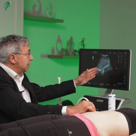
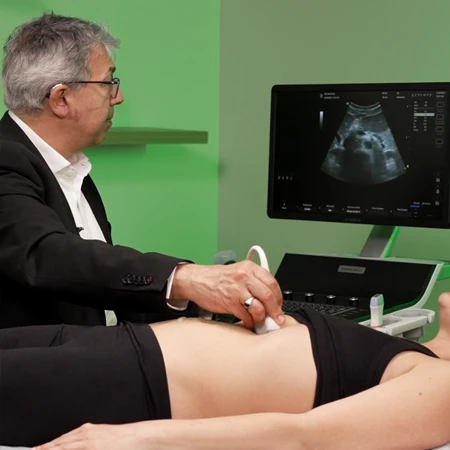
- *NEW* Video format with subtitles, led by Dr. Michel ATHOUËL, a renowned expert in ultrasound imaging
- Downloadable transcript available
- Meets the CE requirements of the following states: California, Texas, Florida, Kentucky, Massachusetts, and New Mexico
- Meets ARRT® CE reporting requirements
- Meets ARDMS® and APCA® CME reporting requirements
- Hassle-free 30-day full refund policy*
Pulmonary ultrasound, once an uncommon practice, has proven its value in modern medical examinations. Initially, ultrasound was not used for organs containing air or gas, such as the lungs and digestive tract. Over time, however, extensive research has highlighted its potential, particularly in detecting pleural effusion and alveolo-interstitial syndromes. Notably, a 2004 study demonstrated the superiority of pulmonary ultrasound over auscultation and chest X-rays in identifying pleural effusion and alveolar syndromes. Similarly, a 2016 study found ultrasound’s specificity in detecting pneumothorax, pleurisy, and alveolar syndrome to be significantly high compared to traditional methods. These findings underscore the substantial role of ultrasound in lung exploration, showing its efficacy in cases of consolidation syndrome, pneumothorax, pleural effusion, and interstitial syndromes.
Understanding the anatomical context of the thorax is crucial for effective pulmonary ultrasound. The thorax houses the lungs, heart, diaphragm, and the bony thoracic cage, which includes the ribs. Bone, being a solid structure, obstructs ultrasound waves, necessitating the navigation of the probe between the ribs. Proper probe orientation is essential, especially given the oblique orientation of the ribs laterally. Despite the complexities posed by the ribcage and scapulae, the lungs’ substantial size requires a methodical approach to exploration, dividing each lung into six parts for comprehensive assessment.
The principles of pulmonary ultrasound emphasize the technique’s simplicity, the challenge of navigating both water and air within the lungs, and the importance of identifying signs and pathologies originating from the pleural line. Additionally, understanding the echo-structural characteristics of the lungs, including solid (bones) and gaseous parts (alveoli, bronchi), and analyzing artifacts such as posterior acoustic shadowing and enhancements, is vital for accurate diagnosis.
| Discipline | Major content category & subcategories | CE Credits provided |
| RA-2018 | Procedures | |
| Thoracic Section | 0.50 | |
| SON-2019 | Procedures | |
| Superficial Structures and Other Sonographic Procedures | 2.25 | |
| RA-2023 | Procedures | |
| Thoracic Section | 0.50 | |
| SON-2024 | Procedures | |
| Superficial Structures and Other Sonographic Procedures | 2.25 |
|
Get it now!
One-time payment. No hidden fees. No extra charges per credit.
|
|
|