
Radiographic Head Procedures
An interactive CE course covering the intricate details of the anatomy and positioning techniques specific to the skull, sinuses, and facial bones.
- Approved by the ASRT (American Society of Radiologic Technologists) for 3.25 CE Credits
- Subscription duration: 365 days from purchase date
- Downloadable transcript available
- Meets the CE requirements of the following states: California, Texas, Florida, Kentucky, Massachusetts, and New Mexico
- Meets ARRT® CE reporting requirements
- Accepted by the NMTCB®
- Hassle-free 30-day full refund policy*
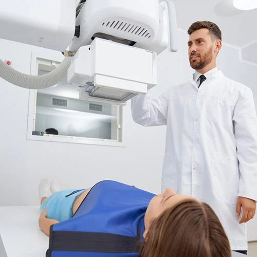
In this comprehensive course, we will delve into the intricate details of the anatomy and positioning techniques specific to the skull, sinuses, and facial bones. We aim to provide attendees with a comprehensive understanding of these critical anatomical structures within the head, along with the specialized positioning required for imaging them accurately. We will start by exploring the unique features of each anatomical structure, highlighting their distinct characteristics and functions within the cranial region. Understanding these features is essential for clinicians and radiographers alike, as it lays the groundwork for precise diagnosis and treatment planning.
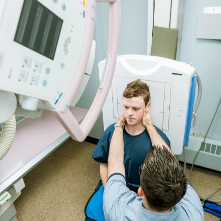
Next, we will delve into the nuances of positioning for imaging the skull, sinuses, and facial bones. This will encompass a thorough examination of patient positioning, equipment setup, and imaging parameters tailored to each specific projection. Attendees will learn the intricacies of positioning techniques, ensuring optimal visualization of the targeted anatomical structures while minimizing artifacts and distortion.
Furthermore, we will establish clear evaluation criteria for assessing the quality of projections obtained from skull, sinus, and facial bone imaging. This will involve identifying key anatomical landmarks, assessing image clarity, and recognizing common artifacts that may affect diagnostic accuracy.
Finally, we will explore a variety of common head pathologies encountered in clinical practice. By discussing these pathologies in the context of anatomical structures and imaging techniques, attendees will develop a deeper understanding of how different conditions manifest in imaging studies. This knowledge will be invaluable for accurate diagnosis, treatment planning, and patient management.
Overall, this course aims to equip participants with the knowledge and skills necessary to confidently perform and interpret imaging studies of the skull, sinuses, and facial bones. By the end of the session, attendees will have a solid foundation in head anatomy, positioning techniques, evaluation criteria, and the identification of common pathologies, enhancing their clinical practice and patient care.
| Discipline | Major content category & subcategories | CE Credits provided |
| CT-2017 | Procedures | |
| Head, Spine, and Musculoskeletal | 1.00 | |
| NMT-2017 | Procedures | |
| Other Imaging Procedures | 1.00 | |
| RAD-2017 | Procedures | |
| Head, Spine and Pelvis Procedures | 3.25 | |
| THR-2017 | Procedures | |
| Treatment Sites and Tumors | 1.00 | |
| RA-2018 | Procedures | |
| Musculoskeletal and Endocrine Sections | 2.00 | |
| PTH-2019 | Procedures | |
| Treatment Sites | 1.00 | |
| MRI-2020 | Procedures | |
| Neurological | 1.00 | |
| CT-2022 | Procedures | |
| Head, Spine, and Musculoskeletal | 1.00 | |
| NMT-2022 | Procedures | |
| Other Imaging Procedures | 1.00 | |
| RAD-2022 | Procedures | |
| Head, Spine and Pelvis Procedures | 3.25 | |
| THR-2022 | Procedures | |
| Treatment Sites and Tumors | 1.00 | |
| RA-2023 | Procedures | |
| Musculoskeletal and Endocrine Sections | 2.00 |
Section 1: The Skull
- Anatomy
- Positioning and procedures
- Pathologies
- Conclusion
Section 2: Facial Bones
- Anatomy
- Procedures
- Pathologies
- Conclusion
Section 3: Sinuses
- Anatomy
- Positioning and procedures
- Pathologies
- Conclusion
|
Get it now!
One-time payment. No hidden fees. No extra charges per credit.
|
|
|
Unlimited CE Credits
For Only $49.99
1 – year Access – Doesn’t Auto-Renew
Guaranteed 30-day Refund Policy


