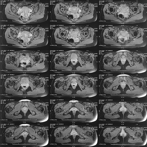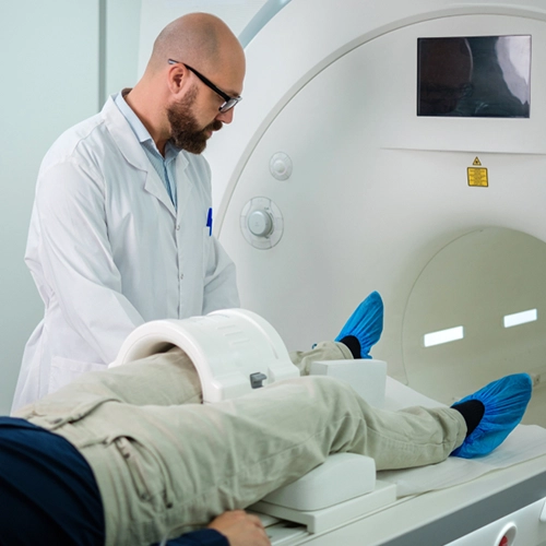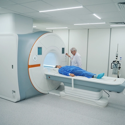

MRI of the Pelvis
Master the fundamentals of MRI pelvis imaging, including protocols for MRI pelvis with and without contrast, anatomy, patient care, and artifact management.
- Approved by the ASRT (American Society of Radiologic Technologists) for 4.00 Category A CE Credits
- Subscription duration: 365 days from purchase date
- Downloadable transcript available
- Meets the CE requirements of the following states: California, Texas, Florida, Kentucky, Massachusetts, and New Mexico
- Meets ARRT® CE reporting requirements
- Accepted by the ARMRIT®
- Hassle-free 30-day full refund policy*
Explore the intricacies of MRI pelvis imaging with this in-depth course tailored for radiologic technologists and healthcare professionals. Gain expertise in planning and performing MRI pelvis with and without contrast while understanding the nuances of imaging protocols for both male and female patients.
This comprehensive course covers the anatomy and physiology of the MRI of pelvis region, providing a solid foundation for understanding the structures being imaged. You’ll learn essential patient care and preparation techniques to ensure comfort and safety, along with detailed insights into imaging sequences tailored specifically for MRI pelvis with and without contrast. The course also provides step-by-step guidance on protocols for the female and male pelvis, addressing unique clinical considerations for each. Additionally, it offers strategies to identify and manage artifacts, explores the safe and effective use of contrast media for enhanced imaging, and emphasizes proven methods to achieve superior image quality.
Whether you are looking to refine your skills or expand your knowledge, this course offers a comprehensive understanding of MRI pelvis, empowering you to deliver accurate and reliable diagnostic imaging.
| Discipline | Major content category & subcategories | CE Credits provided |
| THR-2017 | Procedures | |
| Treatment Sites and Tumors | 0.50 | |
| RA-2018 | Patient Care | |
| Pharmacology | 0.50 | |
| RA-2018 | Procedures | |
| Abdominal Section | 0.50 | |
| PTH-2019 | Procedures | |
| Treatment Sites | 0.50 | |
| MRI-2020 | Patient Care | |
| Patient Interactions and Management | 0.50 | |
| MRI-2020 | Image Production | |
| Physical Principles of Image Formation | 1.00 | |
| Sequence Parameters and Options | 0.50 | |
| MRI-2020 | Procedures | |
| Body | 2.00 | |
| THR-2022 | Procedures | |
| Treatment Sites and Tumors | 0.50 | |
| RA-2023 | Patient Care | |
| Pharmacology | 0.50 | |
| RA-2023 | Procedures | |
| Abdominal Section | 0.50 | |
| MRI-2025 | Patient Care | |
| Patient Interactions and Management | 0.50 | |
| MRI-2025 | Image Production | |
| Physical Principles of Image Formation | 1.00 | |
| Sequence Parameters and Options | 0.50 | |
| MRI-2025 | Procedures | |
| Body | 2.00 |
Section 1: Anatomy and Physiology
- Anatomy and physiology of the female pelvis
- Vascularization
- Physiology
- Anatomy and physiology of the male pelvis
Section 2: Patient Care and Preparation
- Medical history and assessments
- Contraindications
- Patient preparation
Section 3: Sequences
- Introduction
- Fast spin echo
- Gradient echo
- 3D LAVA/3D LAVA flex
- PROPELLER
- DISCO
- Diffusion
Section 4: Female Pelvis Protocols
- Defecography-MRI protocol
- Rectum and anal fistula
- Endometriosis
Section 5: Male Pelvis Protocols
- Prostate indications and protocols
Section 6: Artifacts
- Motion
- Flow
- Aliasing
- Magnetic susceptibility
- Ferromagnetic
Section 7: Contrast Media
- Introduction
- Reminder of T1 and T2 weightings
- Contrast agents
- Indications
- Contraindications
- Side effects
- Contrast administration
Section 8: Image Quality
- Contrast
- Signal-to-noise ratio (SNR)
- Spatial resolution
- Summary
|
Get it now!
One-time payment. No hidden fees. No extra charges per credit.
|
|
|


