
MRI of the Head
Master the essentials of head MRI exams with this ASRT-approved CE course. Designed for radiologic technologists, it covers head anatomy, imaging protocols, artifacts, and common pathologies.
- Approved by the ASRT (American Society of Radiologic Technologists) for 5.75 Category A CE Credits
- Subscription duration: 365 days from purchase date
- Downloadable transcript available
- Meets the CE requirements of the following states: California, Texas, Florida, Kentucky, Massachusetts, and New Mexico
- Meets ARRT® CE reporting requirements
- Accepted by the ARMRIT®
- Hassle-free 30-day full refund policy*
Mastering the fundamentals of MRI of the brain and other head structures is essential for radiologic technologists committed to delivering accurate and high-quality imaging. This comprehensive course provides everything you need to confidently perform an MRI procedure, offering a complete understanding of the procedure from start to finish. The “MRI of the Head” course begins by building a solid head anatomy and physiology foundation, guiding you through the brain, internal auditory canal, orbits, sinuses, and vascular structures. You’ll gain skills to correlate this anatomy with real MRI images across different planes, enhancing your ability to visualize and assess key structures.
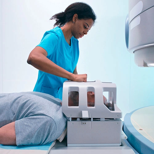
Moving beyond anatomy, the course dives into the clinical indications for ordering an MRI of the brain and other head structures and explores contraindications and special considerations, such as unique approaches for pediatric, geriatric, bariatric, and trauma patients. From there, you’ll learn the essentials of patient preparation, positioning, and the use of surface coils to ensure optimal imaging outcomes. Detailed explanations of imaging protocols for each head structure—including the brain, internal auditory canal, orbit, sinuses, and vascular imaging like MRA and MRV—will teach you how to adapt techniques to account for size and orientation differences.
You’ll also tackle common challenges in imaging by learning to identify and avoid artifacts such as motion, susceptibility, and chemical shift artifacts. The course concludes with an exploration of pathologies, including tumors, vascular abnormalities, degenerative diseases, infections, and trauma, offering insights into their appearance on real MRI images. Whether you’re looking to expand your expertise or refine your skills, this course equips you with the knowledge to perform an MRI of the brain and other head structures with confidence and precision.
| Discipline | Major content category & subcategories | CE Credits provided |
| THR-2017 | Procedures | |
| Treatment Sites and Tumors | 1.00 | |
| RA-2018 | Safety | |
| Patient Safety, Radiation Protection, and Equipment Operation | 0.25 | |
| RA-2018 | Procedures | |
| Neurological, Vascular, and Lymphatic Sections | 2.25 | |
| PTH-2019 | Procedures | |
| Treatment Sites | 1.00 | |
| MRI-2020 | Patient Care | |
| Patient Interactions and Management | 0.25 | |
| MRI-2020 | Safety | |
| MRI Screening and Safety | 0.25 | |
| MRI-2020 | Image Production | |
| Physical Principles of Image Formation | 1.00 | |
| MRI-2020 | Procedures | |
| Neurological | 4.25 | |
| THR-2022 | Safety | |
| Radiation Protection, Equipment Operation, and Quality Assurance | 0.25 | |
| THR-2022 | Procedures | |
| Treatment Sites and Tumors | 1.00 | |
| RA-2023 | Safety | |
| Patient Safety, Radiation Protection, and Equipment Operation | 0.25 | |
| RA-2023 | Procedures | |
| Neurological, Vascular, and Lymphatic Sections | 2.25 | |
| MRI-2025 | Patient Care | |
| Patient Interactions and Management | 0.25 | |
| MRI-2025 | Safety | |
| MRI Screening and Safety | 0.25 | |
| MRI-2025 | Image Production | |
| Physical Principles of Image Formation | 1.00 | |
| MRI-2025 | Procedures | |
| Neurological | 4.25 |
Section 1: Anatomy and Physiology
- Anatomy of the brain
- Anatomy of internal auditory canal
- Anatomy of the orbits
- Anatomy of the sinuses
- Vascular anatomy
- Summary
Section 2: Indications & Contraindications of MRI
- Indications
- Contraindications
- Special considerations
- Summary
Section 3: Preparation and Positioning
- Patient preparation
- Surface coils
- Patient positioning
- Special considerations
- Pediatric patients
- Geriatric patients
- Bariatric patients
- Trauma patients
- Summary
Section 4: Imaging Protocol
- Imaging planes
- Brain
- Internal auditory canal
- Orbit
- Sinuses
- MRA and MRV
- Planning
- Parameters
- Sequences
- Summary
Section 5: Pathologies
- Tumors
- Vascular abnormalities
- Degenerative diseases
- Infections and inflammatory conditions
- Traumas
- Summary
Section 6: Artifacts
- Motion artifacts
- Susceptibility artifacts
- Chemical shift artifacts
- Gibbs (truncation) artifacts
- Wrap-around (aliasing) artifacts
- Flow artifacts
- Partial volume artifacts
- RF interference
- Summary
|
Get it now!
One-time payment. No hidden fees. No extra charges per credit.
|
|
|
Unlimited CE Credits
For Only $49.99
1 – year Access – Doesn’t Auto-Renew
Guaranteed 30-day Refund Policy


