Brain Imaging in Alzheimer’s Disease

In this artcile, we will focus on brain imaging in Alzheimer’s disease, Alzheimer’s MRI technique, and PET scan for Alzheimer’s.


Introduction
Alzheimer’s disease, a progressive neurodegenerative disorder, has become an alarming public health concern as the global population continues to age. Characterized by cognitive decline, memory loss, and a host of behavioral changes, Alzheimer’s poses significant challenges to patients, caregivers, and healthcare providers alike. In the years 2022 and 2023, it is estimated that 1.95% to 1.99% of US citizens were diagnosed with Alzheimer’s disease. This means that about 1 in 53 aged 65 and older in the US are affected. While there is currently no cure for Alzheimer’s, early detection and intervention can significantly improve a patient’s quality of life. Alzheimer’s MRI and PET scan brain imaging has emerged as a crucial tool in understanding, diagnosing, and tracking the progression of the Alzheimer’s disease.
“Alzheimer’s disease, the most common form of dementia, affects more than 50 million people worldwide. It robs people of their independence and is the fifth leading cause of death.” – nature.com
Brain Imaging in Alzheimer’s Disease have revolutionized our understanding of it. By providing non-invasive and highly detailed images of the brain, these techniques enable clinicians and researchers to visualize structural and functional changes associated with Alzheimer’s. There are several key imaging modalities used in the study of Alzheimer’s disease.

Magnetic Resonance Imaging (MRI)
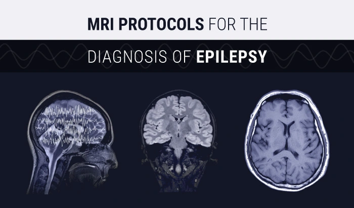
MRI is a widely used imaging technique that provides detailed images of the brain’s structure. Alzheimer’s MRI is particularly valuable for detecting atrophy (shrinkage) in specific brain regions (a hallmark of Alzheimer’s). Alzheimer’s MRI can also reveal abnormalities in brain structure that may contribute to cognitive impairment.
The below image shows progressive atrophy (medial temporal lobes) in an older cognitively normal (CN) subject, an amnestic mild cognitive impairment (aMCI) subject, and an Alzheimer’s disease (AD) subject. Role of structural MRI in Alzheimer’s disease- Prashanthi Vemuri & Clifford R Jack Jr.

https://alzres.biomedcentral.com/articles/10.1186/alzrt47
While there are numerous parameters that can be adjusted in the Alzheimer’s MRI protocol, the following are some of the important parameters and sequences commonly used in brain MR imaging for diagnosing Alzheimer’s disease:
- T1 and T2-Weighted Imaging: High-resolution brain MR imaging T1-weighted and T2-weighted anatomical images are essential for assessing brain atrophy and structural changes for Alzheimer’s disease. Acquisitions are recommended to be performed at high spatial resolution in Alzheimer’s MRI (e.g., 1 mm isotropic voxel size) for onset atrophy detection. 3D MRI provides high-resolution anatomical images of the brain. This high spatial resolution is essential for detecting subtle structural changes that occur in AD (Alzheimer’s disease), such as cortical atrophy and ventricular enlargement. These structural changes are key diagnostic markers
- Fluid-Attenuated Inversion Recovery (FLAIR) Sequences: FLAIR images highlight areas of abnormal signal intensity, often used to detect and quantify white matter hyperintensities. High spatial resolution, long repetition time (TR), and echo time (TE) are adjusted to nullify cerebrospinal fluid signals
- Diffusion-Weighted Imaging (DWI): Measuring the diffusion of water molecules in brain tissue is useful for detecting acute or chronic ischemic changes. multiple diffusion gradient directions may be utilized, as well as different b-values for improved sensitivity
- Functional MRI (fMRI): fMRI measures changes in blood flow and oxygenation in the brain, allowing researchers to investigate how different brain regions are activated during cognitive tasks. It is especially useful for understanding the functional changes that occur as Alzheimer’s progresses. Important techniques to be used while performing a fMRI are Blood Oxygen Level Dependent (BOLD), whole-brain coverage, and appropriate task-based or resting-state paradigms
- Diffusion Tensor Imaging (DTI): DTI is an MRI-based technique that provides insights into the integrity of white matter tracts in the brain. Changes in white matter integrity can be indicative of cognitive decline in Alzheimer’s disease. Important Parameters in Alzheimer’s MRI are to be considered in DTI are high-angular resolution, multiple diffusion directions, and isotropic voxel size (e.g., 2 mm)

Positron Emission Tomography (PET)
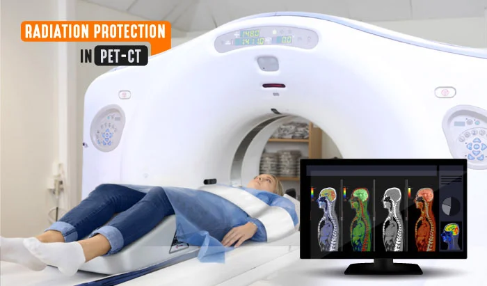
Positron Emission Tomography (PET) scan imaging for Alzheimer’s disease is a valuable tool in its diagnosis and evaluation of progression. When using PET scan for Alzheimer’s diagnosis, several important parameters and techniques are employed to provide useful information about brain function and pathology. Here are some of the key parameters used in PET scan for Alzheimer’s disease:
- Radiotracer Choice: The choice of radiotracer is crucial. For PET scan for Alzheimer’s disease, the most commonly used radiotracers target specific pathological features of the disease. Two primary types of radiotracers are used
- Amyloid Imaging Tracers: These tracers bind to amyloid plaques, a hallmark of AD. Examples include [18F] Florbetapir (Amyvid), [18F] Florbetaben (NeuraCeq), and [11C] PiB (Pittsburgh Compound B)
- Glucose Metabolism Tracers: These tracers measure brain glucose metabolism, which is typically reduced in AD. [18F] FDG (Fluorodeoxyglucose) is the most commonly used tracer for this purpose
-

-
Typical [11C]-Pittsburgh compound B (11C-PIB) positron emission tomography (PET) images of cognitively normal controls (NC), mild cognitive impairment (MCI) cases, and an Alzheimer’s disease (AD) patient. Biomarkers in Alzheimer’s Disease- Book September 2016, Tapan K Khan
- Standardized Uptake Value (SUV): SUV is a measure of radiotracer uptake in a specific region of interest (ROI). In AD, SUV can be used to quantify amyloid plaque burden or changes in glucose metabolism. A decreased SUV in [18F] FDG-PET, for example, is indicative of reduced glucose metabolism, which is characteristic of AD
- Cerebral Blood Flow: Some PET studies may include measurements of cerebral blood flow using tracers like [15O] H2O. In PET scan for Alzheimer’s, reduced blood flow in specific brain regions can be a strong indicator
- Time Activity Curve (TAC): TACs are used to evaluate how the radiotracer accumulates and washes out in different brain regions over time. These curves provide valuable information about tracer kinetics and can help distinguish AD from other conditions
- SUV Ratio: In amyloid PET, the SUV ratio is calculated by comparing the SUV in regions associated with amyloid deposition (e.g., frontal cortex) to a reference region with minimal amyloid deposition (e.g., cerebellum). This ratio helps quantify the amyloid burden
- Region of Interest (ROI) Analysis: Researchers and clinicians define specific ROIs within the brain to measure radiotracer uptake. Common ROIs in AD studies include the frontal cortex, posterior cingulate cortex, precuneus, and parietal cortex
Single-Photon Emission Computed Tomography (SPECT)
SPECT is another functional imaging technique that can provide valuable information about blood flow and brain metabolism. It is sometimes used in conjunction with specific tracers to assess brain function in Alzheimer’s patients.
Early Detection and Differential Diagnosis
One of the most significant advantages of medical imaging in Alzheimer’s research is its ability to detect the disease in its early stages. Early detection is crucial because it allows for timely intervention and the initiation of treatments aimed at slowing the disease’s progression. Furthermore, medical imaging can help differentiate Alzheimer’s disease from other neurodegenerative disorders, such as frontotemporal dementia or vascular dementia, which may have similar clinical symptoms but require different management approaches.

Coronal MRI Brain with Alzheimer’s disease

Sagittal MRI Brain with frontotemporal dementia
Monitoring Disease Progression
Brain imaging is also vital for tracking the progression of Alzheimer’s disease over time. Longitudinal imaging studies can provide insights into how the disease affects the brain’s structure and function, allowing researchers to develop a deeper understanding of the disease’s natural history and potentially identify markers of disease progression.
About 5% of people aged 65 to 74 are diagnosed with Alzheimer’s. Medical imaging is a valuable tool in the research and diagnosis of Alzheimer’s disease. It can help detect the disease in its early stages, which is crucial for timely intervention and treatment, and identify its progression.
Research and Therapeutic Development
The percentage of people with Alzheimer’s disease is increasing with age. In 2022, about 5% of people aged 65 to 74, 13.1% of people aged 75 to 84, and 33.3% of people aged 85 and older have Alzheimer’s disease in the USA. The number of people with Alzheimer’s disease is expected to grow in the coming years as the population ages. By 2060, it is estimated that 13.8 million Americans will have Alzheimer’s disease. Here are some other statistics about Alzheimer’s disease in the US:
- The average age of onset for Alzheimer’s disease is 65 years old
- Women are more likely than men to develop Alzheimer’s disease
- African Americans and Hispanics are more likely than Whites to develop Alzheimer’s disease
- There is no cure for Alzheimer’s disease, but there are treatments that can help manage the symptoms
In addition to its diagnostic and monitoring capabilities, medical imaging plays a pivotal role in Alzheimer’s disease research and the development of potential treatments. By providing detailed data on brain changes associated with the disease, imaging studies aid researchers in identifying new drug targets and evaluating the effectiveness of experimental treatments in clinical trials.
Challenges and Future Directions
While medical imaging has made significant strides in the understanding and management of Alzheimer’s disease, challenges remain. The cost and availability of advanced imaging techniques can be barriers to widespread use, and there is ongoing research to develop more accessible and cost-effective methods for early detection and monitoring.
Furthermore, ongoing research aims to refine imaging biomarkers that can accurately predict a person’s risk of developing Alzheimer’s, ideally before symptoms manifest. This could revolutionize early intervention and prevention strategies.
Conclusion
Medical imaging has transformed our ability to study, diagnose, and track Alzheimer’s disease. By providing valuable insights into the structural and functional changes in the brain, imaging technologies empower clinicians and researchers to understand the disease’s mechanisms better and develop more effective interventions. As technology continues to advance, the role of medical imaging in the fight against Alzheimer’s disease is likely to become even more prominent, offering hope for improved patient outcomes and a brighter future in the battle against this devastating condition.
References
- 2023 Alzheimer’s Disease Facts and Figures. Alzheimer’s Association. [LINK]
- Alzheimer’s Disease. Centers for Disease Control and Prevention. [LINK]
- Basics of Alzheimer’s Disease and Dementia. National Institute on Aging. [LINK]
- Alzheimer’s Disease International. [LINK]
- Alzheimer’s Disease Neuroimaging Initiative: ADNI. [LINK]
- alzres.biomedcentral.com
- Biomarkers in Alzheimer’s Disease- Book September 2016, Tapan K Khan
Disclaimer: The information provided on this website is intended to provide useful information to radiologic technologists. This information should not replace information provided by state, federal, or professional regulatory and authoritative bodies in the radiological technology industry. While Medical Professionals strives to always provide up-to-date and accurate information, laws, regulations, statutes, rules, and requirements may vary from one state to another and may change. Use of this information is entirely voluntary, and users should always refer to official regulatory bodies before acting on information. Users assume the entire risk as to the results of using the information provided, and in no event shall Medical Professionals be held liable for any direct, consequential, incidental or indirect damages suffered in the course of using the information provided. Medical Professionals hereby disclaims any responsibility for the consequences of any action(s) taken by any user as a result of using the information provided. Users hereby agree not to take action against, or seek to hold, or hold liable, Medical Professionals for the user’s use of the information provided.


