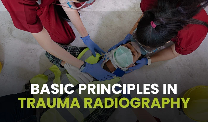
Basic principles in trauma radiography


Improvisation, on the radiographer’s part, is a large component of trauma radiography. The need to know the theory involved in the procedures you perform is essential and the ability to critically think your way through the procedures and make the necessary adjustments to positioning in order to accommodate the condition of the patient is an intricate process. Trauma patients are not “textbook” as I like to refer to patients who continue to be more challenging.
There are some basic rules a trauma radiographer must follow when performing diagnostic radiographic procedures on a trauma patient. The first would include never moving a trauma patient unless there is no alternative. Move the cassette or the central ray. “Take it as it lies”. Bandages, splints, or other immobilization or stabilization devices should never be removed without the assistance of the physician, trauma nurse, or another qualified assistant. If movement of a trauma patient is absolutely necessary, it is essential to obtain adequate assistance to support any injured limbs properly in order to prevent further damage. Also, skeletal injuries always require two views at right angles to each other and long bone radiography require that both joints be included on the radiograph.

A trauma patient most often presents on a back board and is wearing a cervical collar for their protection from movement until proper diagnosis and treatment are administered. Some of the first trauma radiographs that are requested by the trauma surgeon are the AP and cross-table lateral or shoot –thru lateral cervical spine, chest, pelvis (and/or hip) and, on less frequent occasions, an abdomen x-ray. They are performed as follows:
- Cervical Spine – Usually AP and Cross-Table Lateral both on a 10 X 12 image receptor and with the central ray to approximately the level of the fourth cervical vertebra and the x-ray tube angled 15-20 degrees cephalic. The AP is performed at 40” SID while the lateral is performed as close to 72” SID as possible or sometimes in order to visualize C7 better, if you can get it can be performed at 120”SID. This is performed I order to visualize C1 through C7. If C7 still is unable to be visualized then a Swimmers view or CT scan can be performed.
When performing the cervical spine and other radiographs a radiographer is utilizing either the portable x-ray machine or in most cases today’s trauma bays are equipped with x-ray tubes installed as permanent fixtures in the ceiling that move with the same precision as those in a routine radiographic room.
DO NOT REMOVE COLLAR - Chest – If unable to do AP erect chest at as close to 72” SID as possible then a supine chest x-ray is performed at the maximum SID allowable with a 14X 17 image receptor crosswise or lengthwise depending on the patient’s body habitus with CR perpendicular to chest at or as close to T7 as possible. Watch for patient breathing on inspiration. (1)
- Pelvis – Use a 14 X17 grid cassette CW and place your central ray midway between ASIS and symphysis pubis with the top of image receptor 1” above the iliac crest. Utilize a 40” SID and watch for patient’s breathing on expiration.
- Abdomen – With a 14X 17grid cassette LW or CW depending on the patient’s body habitus place the CR at the iliac crest and at the MSP with a 40”SID and watch for the patient’s breathing on expiration.
There are other imaging modalities that are utilized in order to assist in the evaluation and treatment of trauma patients. As indicted previously, when a radiographer is unable to obtain a radiograph that visualizes C7 when performing a cross-table lateral C-spine a CT scan can be used. The majority of trauma patients are taken to CT after the initial evaluation and treatment by the trauma surgeon. The studies vary depending on the patient’s condition. Then he may send the patient directly to the radiology department depending on his/her condition for some additional radiographs. Another modality that may be used is Angiography. This is used when the trauma surgeon wishes to view any vascular defects or abnormalities. Contrast studies would be performed if urinary system evaluation is indicated. Although MRI is the most expensive modality these days, it may give the trauma surgeon much additional information as to the patient’s condition.
In conclusion, each trauma patient is unique and the trauma radiography technologist must evaluate the patient and adapt central ray angles and image receptor placement in order to account for the patient’s injuries, while attempting to reproduce an adequate radiographic image as close to those of routine textbook general radiography as possible. The ability to be able to critically think during each unique trauma case is a crucial aspect to reach this goal. Teamwork, speed, and good communication are other key attributes of a trauma team and successful trauma imaging. The trauma team is all working together for the good of the trauma patient.
References
- Lampignano, J. P., Kendrick, L. E., & Bontrager, K. L. (2021). Bontrager’s textbook of radiographic positioning and related anatomy. Elsevier.
- Long, B. W., Rollins, J. H., Smith, B. J., & Curtis, T. (2019). Merrill’s atlas of radiographic positioning & procedures (Vol. 2). Elsevier.
- Bittle, M. M., Gross, J. A., & Gunn, M. L. (2012). Trauma radiology companion: methods, guidelines, and imaging fundamentals. Wolters Kluwer Health/Lippincott Williams Wilkins.
- Heather Johnson Radiologic Technology Clinical Director at Pima Medical Institute Follow. (n.d.). Trauma Radiography. SlideShare. [LINK]
Disclaimer: The information provided on this website is intended to provide useful information to radiologic technologists. This information should not replace information provided by state, federal, or professional regulatory and authoritative bodies in the radiological technology industry. While Medical Professionals strives to always provide up-to-date and accurate information, laws, regulations, statutes, rules, and requirements may vary from one state to another and may change. Use of this information is entirely voluntary, and users should always refer to official regulatory bodies before acting on information. Users assume the entire risk as to the results of using the information provided, and in no event shall Medical Professionals be held liable for any direct, consequential, incidental or indirect damages suffered in the course of using the information provided. Medical Professionals hereby disclaims any responsibility for the consequences of any action(s) taken by any user as a result of using the information provided. Users hereby agree not to take action against, or seek to hold, or hold liable, Medical Professionals for the user’s use of the information provided.
