Anatomical Variations Found in Cardiac CT


Introduction
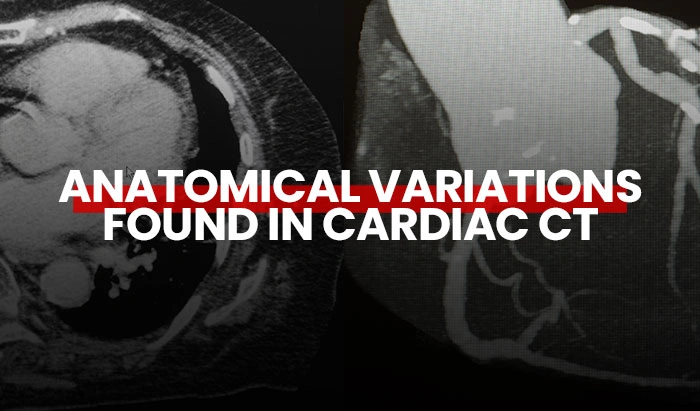
Embarking on our exploration of the intricate marvel that is the human heart, we can’t help but ponder upon a fundamental inquiry: What is this vital organ, and where does it reside within the intricacies of our thoracic cavity? Nestled within the thorax, the heart emerges as a symphony of muscularity, its triangular form gracefully cradled with an apex inclined towards the left, a testament to its dynamic prowess. As we traverse the landscape of cardiac science, cardiac CT imaging emerges as a vital tool, offering detailed depictions of anatomical and physiological variations within this vital organ. Through its lens, we gain insight into the subtle nuances of cardiac morphology and function, unraveling the mysteries of congenital anomalies, coronary artery disease, and other cardiac aberrations that shape the narrative of human health.
Cardiac Anatomy
The heart is made up of four cavities: Two on the right and two on the left with each side having an atrium and a ventricle. You can see the different chambers in this image. Each of the ventricles described has an inlet and an outlet called valves.
Intensity Modulated Radiation Therapy (IMRT)
Intensity-modulated radiation therapy allows for modulating the intensity of the energy of radiation delivered during the session. This technique allows for minimizing radio-induced toxicity, increasing the dose if necessary, and often achieving better curative treatment results. The latest generations of linear accelerators are now equipped with collimators composed of several dozens of thin leaves (multi-leaf collimator, or MLC, composed of 80 to 160 leaves) which allow it to precisely match the shape of the tumor that can be very complex or amorphous. The collimator leaves can also move during irradiation and modulate the flow of the treatment beam. IMRT makes it possible to obtain the desired irradiation at the target tumor volume using successive multibeam techniques, and a very rapid decrease in the dose at its periphery, thus offering better radiation protection of healthy tissue.

Pericardium
In the mediastinum, the pericardium is a sac containing the heart. It consists of a superficial layer known as the fibrous pericardium and a deep layer called the serous pericardium. The pericardium is composed of two layers: The parietal layer towards the outside and the visceral layer towards the inside. These last two layers enclose the pericardial fluid which facilitates the movements of the heart.

Preoperative chest CT shows pericardial effusion. PE: pericardial effusion. https://doi.org
Circulation and Valves
The heart has two circulation systems, the pulmonary circulation, and the systemic circulation. Let us review how the blood travels in the pulmonary circulation, starting with the arrival of blood filled with CO2 through the superior and inferior vena cava.
On the right side of the heart, contraction of the RIGHT atrium propels blood into the RIGHT ventricle through the tricuspid valve. Contraction of the RIGHT ventricle propels blood through the pulmonary valve to the pulmonary arteries. The pulmonary arteries will allow, distally, gas exchanges for the re-oxygenation of the blood. We must remember a particularity here: The pulmonary arteries contain so-called “venous” blood because it is rich in CO2.
By following pulmonary circulation, the oxygenated blood returns to the heart by the pulmonary veins allowing the LEFT atrium to fill up. Again, we note a particularity here: The pulmonary veins contain so-called “arterial” blood because it is rich in oxygen.
The contraction of the LEFT atrium propels blood into the LEFT ventricle through the mitral valve. This oxygenated blood is then propelled into the systemic circulation by contraction of the LEFT ventricle. The left ventricle contraction capacity, called the ejection fraction, must be between 60 and 70 percent. By passing through the aortic valve, this blood enters the aortic artery from which the coronary arteries originate.

Coronary Arteries
There are 2 coronary arteries, the right and left coronary arteries. These arteries supply the heart muscle.
- Right Coronary Artery: The right coronary artery, sometimes called RCA, forms a “C” when arising from the antero-right aortic sinus
- Left Coronary Artery: Known as the common trunk artery, it gives rise to the anterior interventricular artery and the circumflex artery
Coronary CT Angiography
Following the principles of any examination conducted with cardiac gating, standard coronary CT angiography remains an essential basis for many heart studies. The patient’s heart rate will determine the most appropriate technique to adopt.
Slow rhythms allow acquisition in Prospective mode (Prospective mode is an ECG-triggered coronary CT angiography), which will reduce the radiation dose of the exam. Faster rhythms need acquisition in retrospective mode to be able to reconstruct the images as clearly as possible. This will be accompanied by a higher dose. Studies of the heart should include analysis of the atrial and ventricular cavities.

Severe blockage in the right coronary artery on cardiac CT. https://oklahomaheart.com
Main Anatomical Variations
We have already learned that the heart is made up of chambers, valves, and vessels. Anatomical variants are numerous, especially in children.
Valve
Variants of the aortic valve have been widely studied, resulting in many interventions. The tricuspid valve is sometimes bicuspid, with or without raphe (the conjoined area of the fused cusps. Occurring in 1% to 2% of the population, the bicuspid aortic valve (BAV) is the most common congenital cardiac malformation.)

Atrium
The left atrium also attracts a lot of interventions because of its multitude of variations. There are several types of anatomical variants of the implantation of pulmonary veins.
Abnormal pulmonary venous return (APVR) is extremely rare. Total anomalous pulmonary venous return (TAPVR) is a heart disease in which the 4 veins that take blood from the lungs to the heart do not normally attach to the left atrium (left upper chamber of the heart). Instead, they attach to another blood vessel or the wrong part of the heart. It is present at birth (congenital heart disease).

Patent Foramen Ovale
As a baby grows in the womb, an opening called the foramen ovale sits between the upper heart chambers or atria. Normally, it closes on its own within the first few weeks after birth. When the foramen ovale does not close, it is called a patent foramen ovale. A patent foramen ovale (PFO) is a hole in the heart that does not close the way it should after birth and is a small flaplike opening between the atria. Most people never need treatment for patent foramen ovale.


CT scan of a 55-year-old patient with patent foramen ovale. The image here is a coronal oblique, showing a well-defined contrast-enhanced connection between the left and the right atria. https://doi.org
Tetralogy of Fallot
Tetralogy of Fallot is a rare condition caused by a combination of four heart defects that are present at birth (congenital). These defects, which affect the structure of the heart, cause oxygen-poor blood to flow out of the heart and to the rest of the body. Infants and children with tetralogy of Fallot usually have blue-tinged skin because their blood does not carry enough oxygen.
Tetralogy of Fallot is often diagnosed while the baby is an infant or soon after. Sometimes, depending on the severity of the defects and symptoms, tetralogy of Fallot is not detected until adulthood. All babies who have tetralogy of Fallot need corrective surgery.


These images are from a case of a neglected TOF and pulmonary atresia in an adult patient. Images a and b are 3D VRT MDCT images showing innumerous MAPCAs filling the mediastinum arising from descending thoracic aorta as well as right and left subclavian and internal mammary arteries.Images c and d are reconstructed MDCT showing pulmonary atresia and non-confluent right and left pulmonary arteries arising from MAPCAs rather than main pulmonary artery (red arrows). Image e is a reconstructed MDCT showing ascending aortic aneurysm as a result of long-standing volume loading in this patient. https://doi.org
Aortic Coarctation
Coarctation of the aorta is a congenital heart defect. It is also called aortic coarctation. This defect affects the baby’s aorta the largest artery in the body. It carries oxygen-rich blood from the baby’s heart to the rest of the body. When a baby has aortic coarctation, one area of the aorta is narrower than normal. This is one of the most common birth defects representing about 7% of all heart defects. It is two to three times more common in males than in females.
Picture a long balloon that is used to make balloon animals for kids. You twist the balloon at one point to begin forming a shape. This causes the balloon to be pinched inward at that point. The pinch in the middle of the balloon is like what an aortic coarctation resembles. That pinched point might be very narrow and cause severe symptoms soon after birth. Or it might be narrower than normal but wide enough to let blood pass through. In that case, symptoms might not appear until later in childhood or adolescence. Symptoms such as hypertension (high blood pressure) may lead to the detection of aortic coarctation.


This is a CT scan image of a 37-year-old patient with a newly diagnosed aortic coarctation. The curved multiplanar reconstruction image of the coarctation segment shows a discrete shelflike structure (arrow). DOI:10.2214/AJR.14.12529
Surgical Interventions
Understanding the different variations is immensely helpful to clinicians planning interventional procedures to treat coronary artery disease after diagnosis, particularly when there are secondary changes of calcification, plaque formation, and stenosis. Treatments for coronary artery disease can include:
- Balloon angioplasty: A balloon inflated inside the blocked artery to increase blood flow
- Coronary artery stent: A tiny mesh coil expanded inside the blocked artery to keep the pathway open
- Atherectomy: Using a device on the end of a catheter to cut away plaque from the artery wall
- Laser angioplasty: Light used to remove blockages in the coronary artery
- Coronary artery bypass: Grafting a portion of a vein above and below the artery to bypass the blockage. (Veins are usually from the leg but can be from the chest or arm. Sometimes multiple bypass procedures need to be in place to allow the blood to flow fully to the heart
- Ablative procedures: Anatomy of the right atrium is of particular importance when considering interventional procedures including ablation and pacemaker insertion. Cardiac ablation is a procedure that is used to correct heart rhythm problems such as AFib
Summary
Many conditions can affect the heart’s tissue. Cardiomyopathy is when the heart muscle becomes enlarged, thick, or rigid, and as a result, the heart becomes weaker and is less able to pump blood through the body and maintain a normal electrical rhythm. Heart inflammation is inflammation in one or more of the layers of tissue in the heart, including the pericardium, myocardium, or endocardium leading to serious complications, including heart failure, cardiogenic shock, or irregular heart rhythm. And of course, congenital heart disease, when the heart does not develop in the typical way. A congenital heart defect can happen at any point during the development of an unborn baby, or embryo.
We have reviewed how Cardiac CT is an imaging technique that can provide imaging that depicts various anatomical and physiological cardiac variations. It is important to understand normal heart anatomy and physiology to be able to recognize abnormal variations. It is hoped that this article has shown you some of those variations.
References
Understanding the different variations is immensely helpful to clinicians planning interventional procedures to treat coronary artery disease after diagnosis, particularly when there are secondary changes of calcification, plaque formation, and stenosis. Treatments for coronary artery disease can include:
- https://www.drugs.com
- https://www.ajronline.org
- https://www.primalpictures.com
- https://www.nhlbi.nih.gov
- Pericardial tamponade caused by massive fluid resuscitation in a patient with pericardial effusion and end-stage renal disease -A case report – Soonjae Hwang, Ji Young Bae, Tae-Wan Lim, In-Suk Kwak, Kwang-Min Kim
- Department of Anesthesiology and Pain Medicine, Hangang Sacred Heart Hospital, Seoul, Korea – https://doi.org
- Multi-detector computed tomography in the assessment of tetralogy of Fallot patients: is it a must? – https://oklahomaheart.com
- https://doi.org
- CT and MRI of Aortic Coarctation: Pre- and Postsurgical Findings DOI:10.2214/AJR.14.12529
Disclaimer: The information provided on this website is intended to provide useful information to radiologic technologists. This information should not replace information provided by state, federal, or professional regulatory and authoritative bodies in the radiological technology industry. While Medical Professionals strives to always provide up-to-date and accurate information, laws, regulations, statutes, rules, and requirements may vary from one state to another and may change. Use of this information is entirely voluntary, and users should always refer to official regulatory bodies before acting on information. Users assume the entire risk as to the results of using the information provided, and in no event shall Medical Professionals be held liable for any direct, consequential, incidental or indirect damages suffered in the course of using the information provided. Medical Professionals hereby disclaims any responsibility for the consequences of any action(s) taken by any user as a result of using the information provided. Users hereby agree not to take action against, or seek to hold, or hold liable, Medical Professionals for the user’s use of the information provided.
