
Exploring Conventional X-Ray Imaging in Radiography
Explore conventional radiography with Dr. Jean Louis Brasseur in this video course, covering anatomy, pathologies, and imaging techniques for accurate diagnoses.
- Approved by the ASRT (American Society of Radiologic Technologists) for 6.00 CE Credits
- Subscription duration: 365 days from purchase date
- Downloadable transcript available
- Meets the CE requirements of all states including: California, Texas, Florida, Kentucky, Massachusetts, and New Mexico
- Meets ARRT® CE reporting requirements
- Accepted by the NMTCB®
- Hassle-free 30-day full refund policy*
Explore conventional radiography with this comprehensive 25-video course, delivered by world-renowned Dr. Jean Louis Brasseur, a leading expert in osteoarticular imaging. Designed for radiologic technologists and physicians, this course delves into the practical applications of clinical radiology through real case studies and in-depth exploration of anatomical structures.
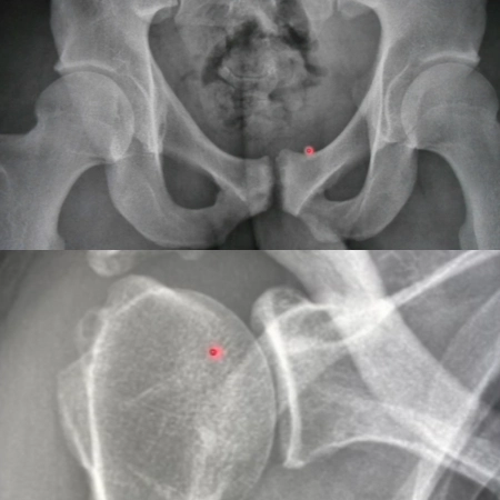
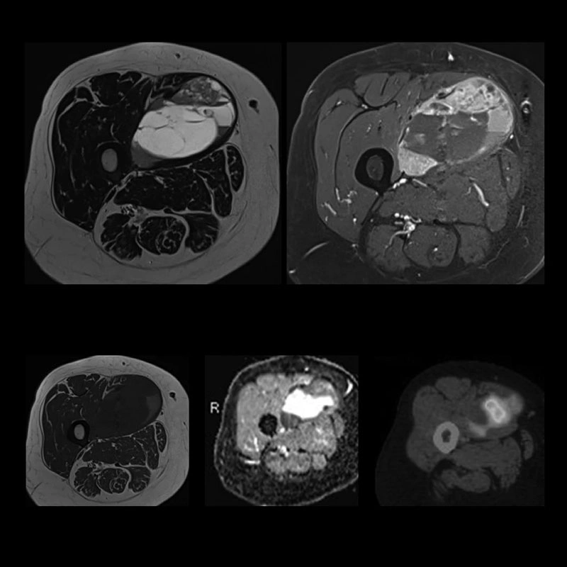
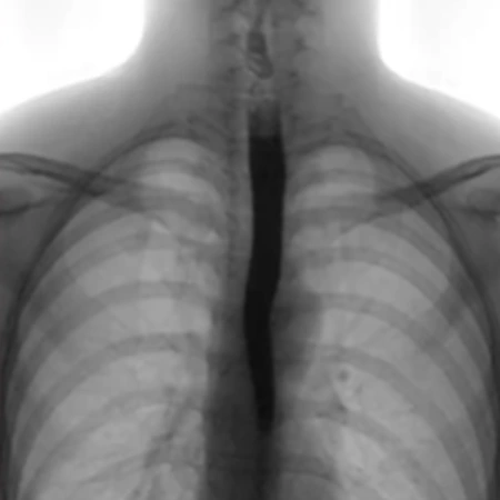
Dr. Brasseur will guide you through the anatomy of the upper extremity (shoulder, hand, and wrist), lower extremity (hip, knee, ankle, and foot), and spine. He will demonstrate how to accurately identify anatomical structures on real X-ray images and explain the visual representation of different pathologies. By showing you various clinical cases, Dr. Brasseur will highlight how different pathologies appear on radiographs, as well as the best positions and techniques for clearly capturing these conditions. He will emphasize the importance of maintaining high-quality imaging to prevent misdiagnosis and ensure accurate interpretation.
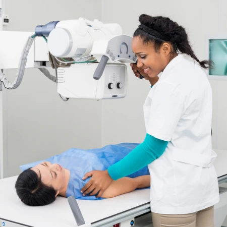
As Dr. Brasseur walks you through these clinical cases, you’ll gain an understanding of the most effective techniques for capturing optimal images. He will show you how positioning can significantly impact the clarity of radiographs, especially when diagnosing specific pathologies. The course will also focus on the role of imaging quality in achieving accurate diagnoses, teaching you how to prevent common pitfalls and errors that can lead to misinterpretation of findings. This comprehensive approach ensures you can confidently apply these skills in clinical practice and provide the highest level of patient care.
With a perfect balance of theoretical insights and hands-on case applications, this course equips you with the skills to expertly analyze conventional radiography images and enhance your interpretation and diagnostic expertise.
This course is not included in the All-Access Pass or packages
| Discipline | Major content category & subcategories | CE Credits provided |
| RAD-2017 | Procedures | |
| Head, Spine, and Pelvis Procedures | 2.00 | |
| Extremity Procedures | 4.00 | |
| RAD-2022 | Procedures | |
| Head, Spine, and Pelvis Procedures | 2.00 | |
| Extremity Procedures | 4.00 |
Section 1: Shoulder (4 videos)
- Video 1 (26min 37sec)
- Video 2 (20min 50sec)
- Video 3 (14min 56sec)
- Video 4 (10min 20sec)
Section 2: Hand and Wrist (4 videos)
- Video 1 (15min 10sec)
- Video 2 (12min 48sec)
- Video 3 (11min 13sec)
- Video 4 (10min 46sec)
Section 3: Knee (4 videos)
- Video 1 (13min)
- Video 2 (18min 14sec)
- Video 3 (14min 35sec)
- Video 4 (12min 57sec)
Section 4: Hip (4 videos)
- Video 1 (10min 48sec)
- Video 2 (13min 43sec)
- Video 3 (13min 10sec)
- Video 4 (12min 11sec)
Section 5: Ankle and Foot (4 videos)
- Video 1 (27min 10sec)
- Video 2 (17min 31sec)
- Video 3 (21min 52sec)
- Video 4 (20min 18sec)
Section 6: Spine (4 videos)
- Video 1 (15min 39sec)
- Video 2 (15min 57sec)
- Video 3 (13min 24sec)
- Video 4 (18min 4sec)
Section 7: Various Cases (1 video)
- Video 1 (11min 19sec)
|
Get it now!
One-time payment. No hidden fees. No extra charges per credit.
|
|
|


